Micro-Focused X-Ray Computed Tomography (microCT / μCT)
MicroCT is a non-destructive imaging technique that uses X-rays to generate high resolution 3D (volume) datasets from samples. Unlike a conventional clinical X-ray system, where image resolution is typically ranging between 0.25 -1.50 mm, this technique can offer spatial resolution down to 6 -8 μm. The scanner comprises an X-ray source, a rotation stage and an X-ray detector. To acquire a tomogram the specimen is mounted on to the rotation stage and is subsequently illuminated by X-rays during a full 360 degree rotation. During this process thousands of radiographs are collected, which are then reconstructed into a 3D data set
Conventionally, X-ray imaging is associated with the examination of hard, high contrast structures like metals, bone etc, but with appropriate choice of detector and source can be adapted for imaging softer tissues.
We have 3 microCT systems:
- A miLabs live animal imaging system (up to medium rabbit size) with both microCT and optical (fluorescence and luminescence) imaging in 3D and 4D, installed 2020. Contact Katie Dexter for more information.
- The first prototype machine of Nikon's Med-X1 series. This experimental / development scanner was designed as a soft tissue imaging research tool for biology and medicine and was commissioned in 2016.
- A larger follow up commercial Nikon Med-XR series machine, again optimised for soft tissue imaging and installed in 2019.
The 2 Nikon systems are run as a X-Ray Histology Facility (XRF) in collaboration with the μ-VIS X-Ray Imaging Centre based in Engineering on the main University Campus, which is part of the National Research Facility for X-ray Computed Tomography (NXCT) and houses multiple complementary scanning systems supporting a wide range of sample sizes and types with imaged volumes up to 1.5 x 1 x 1m, and spatial resolutions down to approximately 200 nm. Both of our Nikon Med-X systems are managed by μ-VIS. Please contact Orestis Katsamenis in μ-VIS for further information.
Find out more about microCT imaging of soft tissues here.
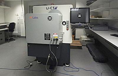
MiLabs 3D/4D optical imaging/microCT system
An integrated, non-invasive, non-destructive imaging system to track the spatial and temporal distribution of tissue structures and physiologically relevant labels in tissue-engineered constructs, and tissue samples in live animals (bone and soft tissues including lung, liver, adipose tissue, vascular systems and cancerous tissue).
- X-ray tomography with a spatial resolution down to ca. 13 μm in each lateral dimension with nominal isotropic voxel resolution down to 2.4 μm.
- 3D fluorescence, bioluminescence and Cherenkov luminescence tomography in the visual to near-IR range (340 nm to 830 nm; up to six emission filters and up to 11 excitation filters; C-MOS detector) with a resolution of ca. 3 mm. Dyes are separated using spectral unmixing.
- ability to scan living organisms up to the size of a small rabbit (up to five mice can be scanned at once).
- correlative imaging to map fluorescence/luminescence images onto X-ray CT images and white light images.
- data manipulation via MiLabs software and/or third party software.
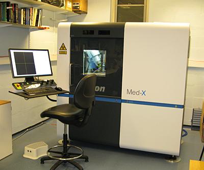
Nikon Med-X Alpha (prototype)
- 130 kVp X-Ray source
- optimised for biomedical imaging
- standard reflection target(~2µm spot)
- 2 x 2k flat panel detector
- samples up to ~300 mm and up to 15 kg
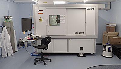
Nikon XT microCT
- optimised for soft tissue imaging
- multi sample loader