
Imaging and Microscopy Centre

More Information

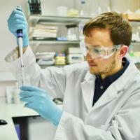
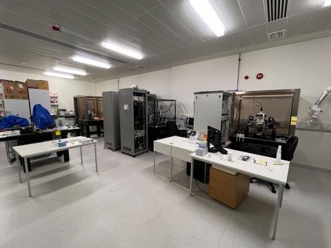
About the Imaging and Microscopy Centre

The Imaging and Microscopy Centre is a core facility for advanced life sciences imaging at Southampton University.

We cater for a broad range of research requirements, from internal university users to industry and other institutions.

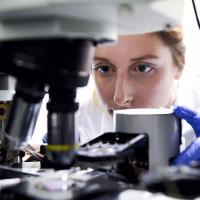
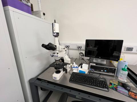
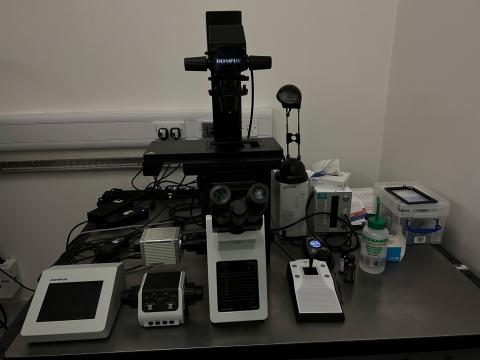
Facilities and equipment

Types of equipment provided by the IMC:

We provide image analysis software for 2D and 3D datasets. You can book our equipment, big-data handling, image analysis and training. We can also supply quotations and assistance for grant applications.

Technical specification

Flow cytometry

We have a flow cytometry machine for measuring cells in solution.

Millipore Guava
Sample format

Single 1.5 ml Eppendorf tube

Filters
Excitation laser and Emission
  • Blue 488 nm (nanometer), green 525/30 nm
  • Red 642 nm, yellow 583/26 nm
  • red 661/15 nm
  • red 695/50 nm
  • forward Scatter
  • side Scatter

Western blotting

We have western blotting equipment for detecting specific proteins in tissue samples

LiCor Odyssey NIR fluorescence flatbed scanner
Detectors

2 x Photomultiplier tube

Lasers and filters

Excitation laser λ and emission filters
  • 685 nm - 700 nm LP (Longpass filters)
  • 785 nm - 800 nm LP

Microinjection

We have microinjection equipment for precision injection very small amounts of fluid into biological cells or nuclei

Eppendorf femtojet electronic microinjection system

Mounted on Zeiss Axiovert 200 stand

Analysis software

Industry-standard image analysis software packages are provided installed on a high-spec analysis PC which can be reserved via the IMC booking calendar:

Imaris – 3D image analysis software

FlowJo – flow cytometry analysis software.

Matlab

FIJI
 

Contact us

Dr Mark Willett

Contact us

Building 85, Highfield Campus, Southampton SO17 1BJ
We’re open Monday to Friday 09:00 to 17:00 UK time.
Back
to top