Skip to main navigationSkip to main content
The University of Southampton
µ-VIS: Multidisciplinary, Multiscale, Microtomographic Volume Imaging

About the Centre

A dedicated centre for Computed Tomography (CT) at Southampton,

providing complete support for 3D imaging science, serving Engineering-, Biomedical-, Environmental- and Archaeological Sciences

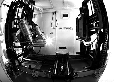
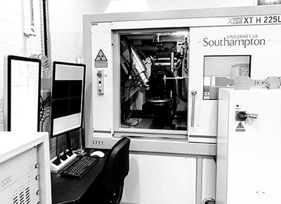
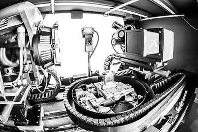
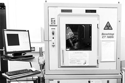
The centre encompasses six complementary scanning systems supporting a wide range of sample sizes with imaged volumes up to 1.5 x 1 x 1m, and spatial resolutions down to approximately 200nm. The various scanners are detailed below. Scanner selection depends on the specimen’s size, material properties such as the density or atomic number of constituent elements, any imposed scan time limitations, as well as the required resolution.

If you have any questions or wish to discuss your needs, please contact us.

Services

µ-VIS offers a range of services to external institutions (academic and industrial) such as:

Equipment

Custom 450kVp / 225kVp Hutch: Nikon/Metris custom design

  • 20-225kV and 100-450kV sources
  • resolutions: ~3µm at low kV, 50µm at 450kV
  • panel and line detectors
  • panel shift system
  • 1x1x1.5m imaging volume
  • 100kg rating
  • temperature controlled
  • large user labyrinth

Custom 225kVp Nikon/Metris HMX ST

  • three configurations
    • transmission (~1µm spot limit, low flux)
    • standard reflection (~3µm spot, 'normal' flux)
    • rotating target (~10µm spot limit, x3-5 flux)
  • 2 x 2k flat panel detector
  • samples to ~300mm and 50kg
  • robotic sample exchange (~150mm height limit)


ZEISS Xradia 510 Versa 3D X-ray microscope

  • 30 -160 kVp
  • 2048 x 2048 pixel detector
  • multiple magnification objectives up to 40x
  • 0.7 µm true spatial resolution
  • phase-contrast imaging modes

160kVp benchtop: Nikon/Metrix CT 160 Xi

  • reflection source, ~3µm spot size limit
  • 1248x1248 flat panel detector
  • samples to ~90mm diameter, 5kg

Gatan XuM/Zeiss Evo MA25 (LaB6 SEM hosted)

  • optimised for 4-12kV operation
  • selection of targets (bulk and thin), ~200nm resolution capabilities
  • phase enhanced contrast
  • 1340x1300 camera
  • samples 1-2mm and below

SkyScan1176 in vivo CT

  • small animal pre-clinical research
  • 100kVp reflection source
  • 11MP camera
  • 9µm in vivo spatial resolutions achievable
  • integrated physiological monitoring
  • bone morphometry tools

Computational facilities

  • GPU-enabled back projection and arithmetic reconstruction packages
  • nVidia Tesla GPU systems enabling high-speed reconstructions
  • dedicated online high-performance storage, live data capacity in excess of 100 TiB
  • dedicated 10 GBit fibre network for fast access to scan data
  • high performance multi-processor workstations with up to 512 GiB RAM for image processing and visualisation
  • software packages include:
    • VG Studio Max
    • FEI Avizo
    • IDL
    • Simpleware (ScanIP, ScanFE, ScanCAD)
    • Digisens DigiXCT
    • Popular open-source tools such as Fiji/ImageJ and MeshLab
Hutch
Custom Nikon/Xtek Hutch (450/225 kVp)
Modified Nikon/Xtek HMX
Modified Nikon/Xtek HMX (225 kVp)
Zeiss/Xradia Versa 510 (160 kVp)
Nikon/Xtek Benchtop (160 kVp)
Privacy Settings