Please find below some examples and case studies of research conducted by the University of Southampton that use animals or utilise animal models.
New bone regeneration from stem cells and bio-materials - From the laboratory to the clinic:
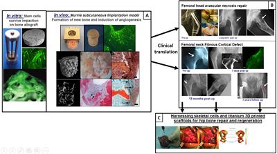
Our skeletal regenerative work is producing candidate bio-materials that support bone stem cell growth and skeletal regeneration improving healing of fractures where major bone loss is a critical clinical problem for the patient. Here we demonstrate in vitro and pre-clinical research using mice on stem cell/bone allograft impaction to regenerate bone and stimulate blood vessel growth (A). This work has led to the clinical translation to repair femoral head avascular necrosis and femoral neck fibrous cortical bone loss (B). Furthermore, this work informed the clinical studies using stem cells in conjunction with 3D printed hips to aid in very large hip bone repair and regeneration (C).
The role of integrins in axonal survival and healing:
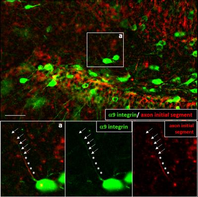
Our work on repairing the central nervous system focuses on traumatic injury to the brain and/or spinal cord. Using rat and mouse models of spinal cord injury, our work has demonstrated that growth-promoting proteins known as integrins are not present in the nerve cells of the mature central nervous system. Our work has demonstrated that a fundamental difference exists in sensory versus motor nerve cells when integrin proteins are re-expressed. Specifically in cell culture models as well as in vivo, sensory nerve cells readily re-express integrin protein throughout the cell leading to successful regenerative growth. Conversely, motor nerve cells in vivo sequester integrin protein within the cell body excluding it from growing tips of the cell resulting in failed regeneration. Several other proteins similar to integrins (transmembrane proteins) have the same issue. Currently we are modifying how integrins are localised within nerve cells using molecular approaches with an aim to increase regenerative growth in motor systems as well as to improve translation of therapeutics into in vivo models.
Coral fluorescents:
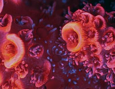
Our in-depth studies of fluorescent coral pigments yielded proteins that can be used as molecular markers in biomedical imaging applications. The pigments can be applied to monitor the molecular workings of living cells and organisms and teach us how diseases work. One these pigments, the red fluorescent protein mRuby from a coral reef-dwelling sea anemone was licensed 2018 for integration in life science products for research purposes. We have discovered and isolated the fluorescent protein “EosFP” from the brain coral Lobophyllia hemprichii which can change its colour from green to red. This protein has subsequently helped the scientist Eric Betzig to develop a novel microscopy technique that enables to study cells with superresolution. An invention for which he was awarded the Noble Prize in Chemistry in 2015. We have used the relevance of coral pigments for medical research to educate the general public about the importance of protecting coral reefs through communicating the value of reefs as source of novel tools and drugs, using the fascinating phenomenon of coral fluorescence as vector to deliver our message. A video on the applications of our research on coral fluorescence can be watched here.
Solent oyster restoration project:
Native European oysters (Ostrea edulis) are a keystone species in the ecology of estuaries and coastal waters and designated as a BAP (Biodiversity Action Plan) species by the UK government. The Solent oyster population, which used to be the largest self-sustaining population in the UK, has been seriously depleted by a mixture of disease, human exploitation and predation in the past 20 years. The School of Ocean and Earth Sciences (SOES) at the University of Southampton is one of the partners in local initiatives, many led by the Blue Marine Foundation, to restore the Solent oyster to its former abundance. SOES research focuses on establishing the physiological and reproductive reactions of oysters to a variety of environmental influences and so establish where, ideally, oysters should be relaid to produce larvae that will settle within the Solent and form the basis for a self-sustaining population in the future. This work combines a mixture of aquarium investigations and field trials.
Understanding disease processes in crustacean aqua-culture:
The Marine Invertebrate Physiology and Immunology (MIPI) Lab have broad interests in how organisms respond to changing marine environments; in particular how predicted climate change scenarios and anthropogenic pollution in the marine environment might impact the health and performance of shellfish species. The group work across a range of scales within organisms, from studies of changes in gene expression in tissues and cells, to studies of metabolism and tissue biochemistry through to assays of whole organism physiology and behaviour.
In a key current project with eight other international partners, and funded through the UK Newton Global Research Partnership, we are investigating the environmental controls on the incidence of disease outbreaks in Asian shrimp and fish aquaculture (Poverty alleviation through control of disease in Asian Aquaculture (PACONDAA)). This project is seeking to understand the environmental controls of global disease pandemics within the shrimp farming industry; diseases which present a significant challenge to the provision of Global Food Security through aquaculture, as the world population approaches an estimated 9.3bn by 2050.
At the University of Southampton, much of our work is based on the immune system of the European shore crab Carcinus maenas as a model species. Blood samples from these crabs are taken to better understand their response to infection and disease resistance. Blood sampling is sometimes carried out after sub-lethal infection with opportunistic pathogenic marine bacteria to confirm our in vitro findings with in vivo infection processes.
Whole Animal Imaging:
Please find a video produced by the University of Southampton's Biomedical Imaging Unit (BIU) on how they safely image animals in a research laboratory.
Non-Technical Summaries (NTS):
Links can be found below to some NTS reports, intended for the public, that have been written by some of our researchers here at the University of Southampton.
NTS reports from expired Project Licences (PPLs)
NTS - Experimental Necrotising Enterocolitis
NTS - Rat Osterolateral Spinal Fusion Model
NTS - Chorioallantoic Membrane Model for Tissue Engineering
NTS - Nervous System Injury and Repair