Confocal microscopy
This uses a pinhole in the light path to discard signal from out of focus regions of a fluorescently labelled (or reflective) sample, thereby generating a sharply focussed, optical section from the sample, without having to physically section it. It is a high magnification, high resolution imaging method which uses a scanning laser beam to excite the fluorescence. Refocussing the microscope and reimaging allows 3D datasets to be collected. Our Leica confocal systems have true spectral detection allowing the simultaneous recording of 4, non-overlapping colour ranges with infinite flexibility and nanometre precision in terms of wavelength detection range.
View a gallery of our confocal microscopy images here.
Lightsheet microscopy
Otherwise known as selective plane illumination microscopy (SPIM), our lightsheet system is a lower magnification, lower resolution method for producing optical sections and 3D images from larger samples (up to 10 x 10 x 5mm)*. It uses a wide, thin sheet of laser light entering the sample from the side (or from multiple sides) to excite fluorescence at a single focal plane which is imaged using a high resolution, ultra sensitive scientific camera. Moving the sample through the laser beam allows signal from different levels within the sample to be captured and 3D data sets generated. Samples for light sheet microscopy often need considerable processing to render them optically clear.
*There are many different types of lightsheet system, using different configurations of light sheet numbers and orientation, providing different degrees of resolution.
Find out more about lightsheet microscopy here.
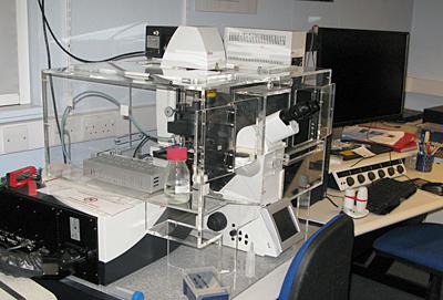
Leica TCS-SP8 Laser Scanning Confocal Microscope
- automated, inverted Leica DMI-8 microscope frame
- Leica LAS-X software control
- 8 laser lines (405nm to 633nm)
- AOBS and AOTF beam path control for fluorescence and reflectance confocal imaging
- 4 spectral detectors
- transmitted light detector with brightfield, DIC, polarising and phase contrast imaging (not confocal but can be superimposed on confocal images)
- objective range x10 to x100 with optical zoom
- glycerol x63 objective for deeper imaging
- electronic stage with relocation, tile scan and navigator functions
- advanced 3D rendering module
- DAPI / FITC / TRITC / CY5 compatible filter cubes for standard fluorescence imaging
- environmental chamber and 5% CO2 in air supply
- camera port
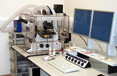
Leica SP5 Laser Scanning Confocal Microscope
- automated, inverted Leica DMI600 microscope frame
- Leica LAS-AF software control
- 9 laser lines (405 to 633 nm)
- AOBS and AOTF beam path control for fluorescence and reflectance confocal imaging
- 4 spectral detectors
- up to 8192 pixel linear resolution
- transmitted light detector with brightfield, DIC, polarising and phase contrast imaging (not confocal but can be superimposed on confocal images)
- objective range x5 to x100 with optical zoom
- glycerol x63 objective for deeper imaging
- 8,000Hz resonant scanner for fast imaging
- FRET and FRAP software wizards
- environmental chamber and 5% CO2 in air supply
- electronic stage with relocation and tile scan function
- DAPI / FITC / TRITC compatible filter cubes for standard fluorescence imaging
- camera port
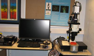
LaVision Light Sheet Ultramicroscope
- Olympus MVX10 microscope frame
- LaVision IMspector software control
- 5 laser lines: (405nm to 785nm)
- single or dual sided illumination
- Andor Neo high sensitivity sCMOS camera
- Olympus MVPlanapo 2XC lens with dipping caps
- 0.63 to 6.3 zoom on microscope stand
- sample size up to 10x10x5mm
- optically clear samples in DBE, water or "clarity"
- live imaging potential for specific sample types