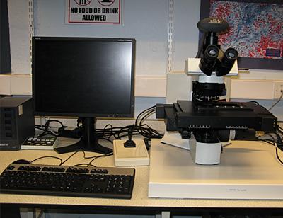
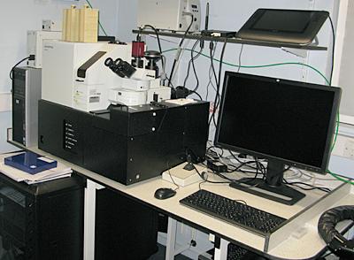
Think Google EarthTM for microscope slides! Virtual microscopy systems allow you to scan an entire slide or selected regions of a slide at a chosen magnification and then zoom in and out and navigate around the scanned slide. Scans are stored as individual image tiles which are assembled / montaged on the fly for low band width, high resolution viewing. Our systems are equipped with adjustable sensitivity tissue content recognition and manual override. Our high throughput system can combine 4 channel fluorescent imaging with brightfield or phase contrast imaging (selected magnifications only for phase contrast).

Olympus dotSlide Virtual Microscopy System (x2)
- Olympus BX51 microscope frame with 4 place slide holder
- automated slide scanning / virtual microscopy system with ultra-high quality, colour Olympus CC12 microscope camera
- brightfield imaging
- objective range x4 to x60
- slide holder for single 3x2 inch slides
- tissue microarray module for assembly of tiles from single cores of TMAs into single images

Olympus VS110 high throughput Virtual Microscopy System
- Olympus BX51 microscope frame with 100 place robotic slide loader (3x1 inch slides only)
- Olympus VS software control
- automated slide scanning / virtual microscopy system with bright field, phase contrast and fluorescence imaging and scanning
- high quality, Allied Vision Technology colour, microscope camera for brightfield and high quality Olympus XM10 monochrome camera for fluorescence
- brightfield imaging, phase contrast (x10 only) and fluorescence imaging
- objective range x2 to x40
- DAPI / FITC / Cy3 / Cy5 filter sets (with quad band dichroic and fast excitation filter wheel)
- Olympus VS desktop software (10 user network licence) for remote analysis of scanned slides
- Z series, Extended Focus Imaging and fluorescence maximum projection options
- scan to database option