Scanning electron microscopy uses a beam of electrons to scan across the surface of a sample and image the surface topography at high resolution and with a large depth of focus. Conventionally, the electron beam interacts with the surface of the specimen (which has been coated with an ultrathin layer of metal) to release secondary electrons which are detected to create the image although backscattered (reflected) electrons from the scanning beam can also be detected. The electron beam also generates x-rays from the sample which can be detected and characterised to identify and map the elemental composition of the sample. Most imaging is done using specially dried and coated samples under high vacuum but low vacuum and environmental modes can be used on some uncoated samples.
View a gallery of our scanning electron microscopy images here.
Our ultra high resolution FEI Quanta 250 SEM is equipped with a Gatan 3View II serial block face imaging system for 3D imaging of samples. Find out more about serial block face imaging here.
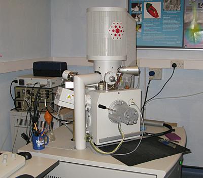
FEI Quanta 250 Scanning Electron Microscope with Gatan 3view2XP serial block face imaging
- ultra high resolution SEM
- 30 Kv / 100,000x magnification
- field emission gun
- high vacuum, low vacuum and environmental modes
- secondary electron and concentric ring backscatter detectors
- digital imaging with auto montage facility
- beam deceleration to enhance surface imaging
- Gatan 3View2XP automatic serial block face imaging down to 25nm slices and near TEM resolution (<2nm)
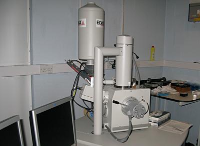
FEI Quanta 200 Scanning Electron Microscope
- 30 Kv / 100,000x magnification medium resolution SEM
- high vacuum, low vacuum and environmental modes
- secondary electron and backscatter detectors
- Oxford Instruments X-Max X-ray microanalysis
- digital imaging to 3584x3094 pixels
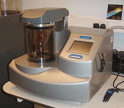
Scanning Electron Microscopy Sample Processing
- processing laboratories with fume cupboard, balance, sample rotators, etc.
- Balzers CPD 030 critical point drier
- Polaron E5100 sputter coater
- Quorum Q150T ES carbon and sputter coater