Transmission electron microscopy shoots a beam of electrons through an ultrathin sample (conventionally a very thin slice of resin-embedded material that has been stained with heavy metals like osmium, lead and uranium). Different components within the sample take up different amounts of the stain and so impede the electrons to different extents, causing contrast in the detected image. The electron beam can also be used to generate X-rays from the different chemical elements in unstained samples. Each element gives off a characteristic spectrum of X-rays so by analysing the spectrum we can map the elemental composition of the sample.
View a gallery of our transmission electron microscopy images here.
.png_SIA_JPG_fit_to_width_INLINE.jpg)
Hitachi HT7700 Transmission Electron Microscope
In 2017, and after 30 years of faithful service, we replaced our Hitachi H7000 TEM with a brand new HT7700 system.
- 120 kV / 600,000x magnification
- digital imaging to 16MP (Morada G3)
- tomography enabled
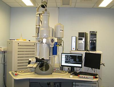
FEI Tecnai T12 Transmission Electron Microscope
- 120 kV / 500,000x magnification
- tilting specimen holder / goniometer stage
- EDAX X-ray microanalysis
- digital imaging to 11MP (Morada G2)
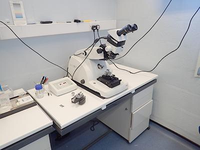
Transmission Electron Microscopy Sample Processing
- processing laboratories with fume cupboard, balance, sample rotators, embedding ovens etc.. (because of the toxicity of many of the chemicals used in TEM processing, all sample prep is done in our laboratories
- 5 x Leica UC7 ultramicrotomes (new 2021), one with live camera for group training and outreach
- 2 x LKB knife makers
- separate processing and ultramicrotomy facilities for diagnostic samples