Ultra-high speed imaging promises new advances in bone fracture modelling
Mechanical engineers at the University of Southampton are improving predictive models for bone fracture risk by tapping into imaging techniques that can analyse impacts at more than a million frames per second.
Researchers in the Materials and Surface Engineering and Biomedical Engineering groups will use the advances to form computer simulations that tackle a number of bioengineering problems, such as predicting injuries in car accidents, understanding hip fractures in falls by the elderly and designing improved body armour in the military.
Dr Lloyd Fletcher is spearheading the three-year programme in a new fellowship from the Leverhulme Trust as he extends methods devised from recent research projects on the mechanical properties of bone and impact testing using ultra-high speed imaging.
“Most existing data that is used to predict bone fracture comes from engineering tests that are performed at very slow loading rates, however many fracture events that occur in real life are the result of impact loading such as falls or car accidents,” Lloyd explains. “If we want to be able to predict fracture risk under impact loading we need models that are informed by experimental data taken under dynamic loading conditions.”
The early career fellowship is developing new test methods that will initially be used on small samples of bone. Data collected from these samples will then be used to develop a numerical model for predicting the breaks in bones.
Building on these advances, the research will use its model in computer simulations of whole bone structures such as a skull or femur. Impact experiments will then be conducted on whole bones and the imaging data compared to the computer simulation predictions.
“There are several key challenges when testing materials under impact loading, including accurate deformation measurement over the whole sample with a high enough sampling rate to capture the whole impact event,” Lloyd says. “Previous technology only allowed for a few point measurements which severely limited the types of tests that could be performed. Recently, cameras have been used to track deformation using image correlation techniques. Coupling these with advances in ultra-high speed imaging has opened up completely new methods which can be used to understand impact loading of materials in unprecedented detail.”
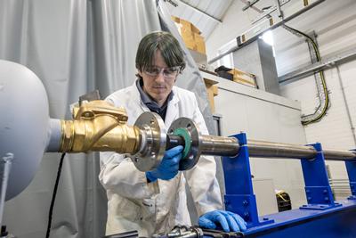
The research is utilising facilities from the University’s Testing and Structures Research Laboratory and the Applied Biomechanics Laboratory, which include a Shimadzu HPV-X camera capable of imaging at to five million frames per second with sufficient image quality for deformation measurement.
Lloyd is working alongside Dr Frances Davis, who is understanding the properties of soft tissue and tendon under impact loading, and Professor Fabrice Pierron, whose Surgery enabled by ultrasonics Programme Grant is developing the next generation of ultrasonic surgical cutting tools. Models created within the Leverhulme Trust fellowship will help inform how these ultrasonic tools will respond when used to cut bone.