Research project: The network in your head: discovering data highways in the human brain
Currently Active:
Yes
To understand how the human brain works, we first need to understand how different parts of the brain are connected. To study this question, scientists are using advanced functional Magnetic Resonance Imaging (fMRI) technologies, which offer a unique window into the operation of the living human brain. Data acquired whilst a person is at rest (i.e. not performing any specific task), allows us to study many aspects of the way in which the human brain is organized and thus enables neuroscientists to study the human brain as a complex, interconnected system. This research is leading to a better understanding of many neural disorders and over the coming years has the potential to significantly contribute to improvements in the diagnosis and cure of many brain diseases.
Project Overview
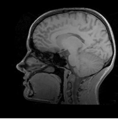
Imaging the human brain
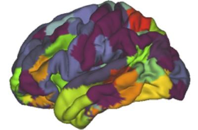
Finding functional brain areas
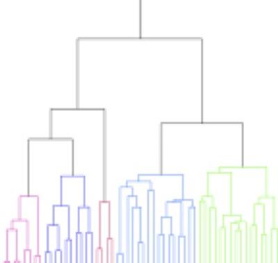
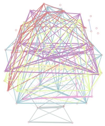
The human brain is organised into functionally distinct regions that are assumed to perform specialised information processing tasks. This neurobiological property of functional specialisation and segregation is complemented by the need to exchange information between different areas. The pattern of this information exchange forms a complex network of functional connections.
Using fMRI data acquired at rest, we develop novel algorithms that are able to estimate the functional divisions in the brain. Based on these estimates, it is then possible to study the information exchange between regions and to build detailed network models of the system level information processing mechanisms that exist in the human brain.
Estimate of functionality
These techniques complement other techniques used in modern neuroscience, such as postmortem studies of cytoarchitecture, in vivo studies of myelin content or methods that look at brain connections using diffusion MRI and tractography. They thus contribute significantly to a deeper understanding of fundamental neural mechanisms of the healthy human brain and provide invaluable information on the things that go wrong in many neural disorders.
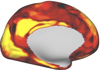
Reliability of the estimates
Estimate of information
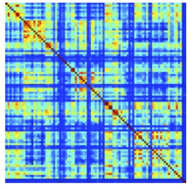
Hierarchical model
Impact
Many neurological disorders such as autism, schizophrenia and epilepsy are now assumed to be directly linked to failures in brain connectivity. A better understanding of normal human brain connectivity and connectivity changes ocuring during these diseas will thus provide important new insights and ultimately lead to better diagnosis and cures. Changes in connectivity have also been shown to act as bio-markers for many degenerative neurological conditions, including alzheimers. The development of advanced techniques for the measurement of brain connectivity thus has the potential to significantly contribute to the development of the early detection of these conditions, long before other symptoms become aparent and, importantly, before irrepairable neural damage occurs.
Part of this work was undertaken for the human connectome project (NIH grant 1U54MH091657-01). Additional funding was provided by EPSRC grants:
EP/J005444/1 Advanced FMRI acquisition, reconstruction and signal processing for dynamic brain network imaging
EP/K037102/1 Constrained low rank matrix recovery: from efficient algorithms to brain network imaging
Publications
Blumensath T,Glasser MF,Jbabdi S, Van Essen DC, Ugurbil K,Behrens TEJand Smith SM, "Spatially constrained hierarchical parcellation of the cerebral cortex with Resting-state FMRI," NeuroImage, 76(1), pp.313-324, Aug. 2013
Blumensath T, Behrens TEJ and Smith SM; "Resting-state FMRI single subject cortical parcellation based on region growing," in Proc. of MICCAI'12, Nice, France, 2012