A new breakthrough in understanding how cells work
Pioneering research at the University of Southampton will help us understand more about the human body at the molecular scale. A new technique developed by Dr Sumeet Mahajan and his group at the Institute for Life Sciences (IfLS) will enable scientists to identify different types of stem cells so they can use the right ones in treatments. It can also help deliver and monitor whether drugs reach the right places in the body and inside cells and tell if they are working.
Pioneering research at the University of Southampton will help us understand more about the human body at the molecular scale. A new technique developed by Dr Sumeet Mahajan and his group at the Institute for Life Sciences (IfLS) will enable scientists to identify different types of stem cells so they can use the right ones in treatments. It can also help deliver and monitor whether drugs reach the right places in the body and inside cells and tell if they are working.
The results of his work, funded by the Engineering and Physical Sciences Research Council (EPSRC), have been published in the influential journal Nano Letters. He is already collaborating with major pharmaceutical companies on its further development.
Stem cell therapy could hold the key to tackling many diseases. They develop into all the various kinds of cells needed in the body - blood, nerves and organs such as the heart, but it is almost impossible to tell them apart, even with the most advanced microscopes, without complex techniques. Up to now, scientists have used intrusive fluorescent ‘markers’ to tag molecules and track each cell but this can alter or damage them and render them useless for therapeutic purposes.
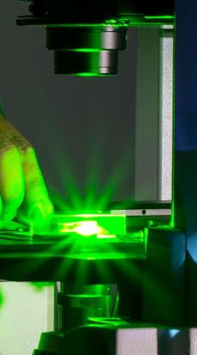
Dr Mahajan uses very tiny 40 nanometer particles of gold, less than 1000th of the width of a human hair, as ‘nanoprobes’ to enter cells. Linked with a natural peptide to avoid rejection, they enhance the observation of the natural vibrations of molecules within the cell and make this motion, otherwise almost invisible, easily detectable with light.
There are many practical applications of this research. For example, some scientists studying Alzheimer’s and Parkinson’s Disease believe replacing the patient’s depleted dopamine generating cells with relevant stem cells or cell grafts may be an effective treatment, but they must be sure they are the right ones to avoid complications and fatalities. Using nanoprobes to gain new insights into the internal activity of cells can also show how drugs are working. Another application is the detection and treatment of cancer cells.

How marine bacteria manage limited ocean i...
The sea is the largest ecosystem on earth, and it harbours two phot...
Read MoreResearch could unlock more precise prognos...
Neuroblastoma study identifies new subgroups with distinct prognose...
Read MoreHigh speed protein movies to aid drug desi...
Researchers from the University of Southampton have developed techn...
Read MoreUp to now, scientists have used intrusive fluorescent ‘markers’ to track each cell but this can alter or damage the cells and render them useless for therapeutic use. By using SERS, we can use very tiny particles of gold, less than 1000th of the width of a human hair, as ‘nanoprobes’ to enter cells.