Imaging Seminar Series Event
- Time:
- 16:00
- Date:
- 14 November 2013
- Venue:
- Room 2209 Life Sciences Building 85 Highfield Campus
For more information regarding this event, please telephone Dr Sumeet Mahajan on 023 8059 3591 or email S.Mahajan@soton.ac.uk .
Event details
Multi-dimensional fluorescence imaging: for cell biology, high content analysis and label-free tissue imaging
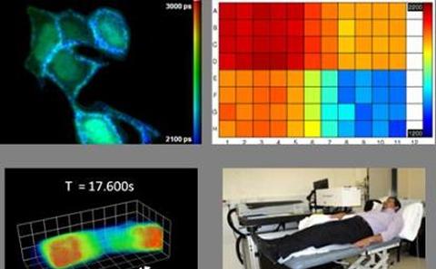
This talk will review our work on quantitative fluorescence microscopy techniques, with an emphasis on fluorescence lifetime imaging (FLIM). FLIM can provide readouts of variations in the local molecular environment of fluorophores, which may be exploited using probes sensitive to e.g. calcium transients or membrane lipid order, or it may be used to sense the proximity of other fluorophores via Forster resonant energy transfer (FRET). Because FLIM is an inherently ratiometric technique that is relatively insensitive to probe concentration or signal attenuation by the sample ("inner filter" effect), it is increasingly applied to read out FRET of appropriately labelled protein-protein interactions to study cell signalling networks.
For cell biology we have developed FLIM microscopes to study fixed and live cells, including high-speed optically sectioned FLIM microscopes for rapid 3-D imaging of cell biology. This has been extended to multiplexing of two independent readouts using FLIM. For drug discovery to take more advantage of FLIM and FRET, e.g. to screen for protein interactions, we have developed automated optically sectioned multiwell plate readers that can image a 96 well plate in less than ~15 minutes, including all sample translation, focussing and FLIM steps. Together with the associated analysis software, we believe that this technology makes FLIM a practical tool for high content analysis (HCA) including for live cell assays. We have recently used this technology to measure dose response curves in an exemplar assay reading out aggregation of the Gag protein in the presence of an inhibitor.
We are also developing a high-speed microscopy technique called oblique plane microscopy (OPM) capable of performing real time 3D imaging of live cells. This method allows us to study events such as calcium sparks and waves in cardiac myocytes in both 2D and 3D at high speed.
For clinical applications, FLIM and MDFI can be applied to autofluorescence to provide label-free molecular contrast in biological tissue and we are applying this to ex vivo and in vivo studies of skin cancer.
Speaker information
Dr Christopher Dunsby,Imperial College London,Department of Physics, Faculty of Natural Sciences