Characterising lung structure with topology
Chronic obstructive pulmonary disease (COPD) is a progressive lung disease resulting in loss of lung function which affects more than 200 million people worldwide. COPD is the fourth leading cause of death worldwide and is likely to become the third leading cause of death by 2020, which places a substantial burden on global health resources. Advances in Computed Tomography (CT) have created opportunities for new diagnostic techniques to enable early discovery of the disease and a more accurate prediction of its severity and prospects and have substantial advantages over spirometry-based diagnostic methods. This, however, creates a pressing challenge to develop new quantitative features obtained by powerful data-characterisation algorithms applied to CT images to uncover characteristics that will have significant medical applications.
Answering this challenge, an interdisciplinary team of mathematicians, clinicians, and image specialists at Southampton University has devised a novel methodology, employing topological data analysis to create new topology-based radiomic features to provide a flexible, computable characterisation of COPD. This work was carried out by Belchi, Pirashvili (Mathematics), Bennett, Conway (Health Sciences) and Djukanović (Medicine), led by Brodzki (Mathematics), and was supported by an EPSRC grant Joining the Dots: From data to insight EP/N014189/1.
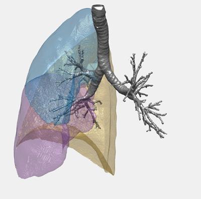
Topology is a part of mathematics designed specifically for the study of complex shapes and in the context of this study it provides an ideal way to develop detailed numerical descriptors of the three-dimensional structure of the lung derived from the CT scans. The research team has demonstrated that these new topological characteristics are powerful and precise enough to provide accurate characterisation of severity of COPD in an algorithmic way. The work, described in an article just published in Scientific Reports introduces a new approach to diagnosing COPD from CT scans and paves the way for large scale studies to turn this method into a breakthrough diagnostic tool be used by clinicians.
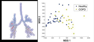
Topological representation of each scan (left) leads to quantitative topological characteristics which allow comparisons between patients. In the picture on the right, each dot represents one CT scan and the proximity of points indicates structural similarity of the lungs. We see a clear separation of healthy controls from COPD patients. The two healthy points inside the COPD region on the right of the picture are healthy smokers, suggesting an undiagnosed problem.