Southampton blood vessel image recognised in national competition
An image of blood vessels in the brain taken by Research Assistant and part time PhD student Matt MacGregor Sharp came runner up in the Reflections of Research competition run by the British Heart Foundation (BHF).
The annual competition aims to reflect the ground-breaking BHF funded research through images.
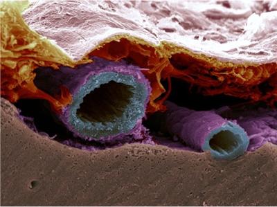
Matt’s image, entitled Subarachnoid Vessels, was taken using a powerful scanning electron microscope and shows a normal artery at the surface of the brain, in stunning detail.
Sitting above the brown brain tissue, the artery appears blue, and its surrounding layer, the pia mater, is shown in purple.
The subarachnoid vessels supply blood to the brain and also act like a drain to remove toxic waste products.
The accumulation of toxic proteins in the walls of arteries and arterioles that supply the brain is known as cerebral amyloid angiopathy (CAA), a key feature of Vascular Dementia.
With ageing, these channels become blocked and waste products accumulate causing disruption and damage to the vessel wall. This image shows a normal artery at the surface of the brain which is susceptible to CAA and subsequent bleeding into the subarachnoid space, a feature known as subarachnoid haemorrhage.
Matt said: “I am delighted to have come runner up in this special competition. These tiny blood vessels have a very important job. To lose the ability to eliminate waste from the brain may lead to vascular dementia, a devastating condition that can affects a lot of people. Understanding the underlying system of this process could bring us a step closer to the design of efficient therapies for vascular dementia.”