New methods for the assessment of human airway ciliary function
New researchers in respiratory medicine have now got access to clear and helpful advice on ciliary function analysis, thanks to a new paper from a Faculty of Medicine scientist.
Ciliary function analysis is key when doctors are investigating airway infections and disease and the deciding what treatments to give the patient.
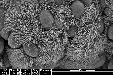
In involves using bespoke high-resolution, high-magnification, high-speed video microscopy analysis to observe and describe ciliary function.
Cilia are moving ‘hair-like’ structures that line the surface of body tissue and in the airway, where they beat in an organised manner to clear mucus and trapped inhaled pathogens or particulates out from the lungs.
Ciliary function analysis is an important tool for helping to diagnose primary ciliary dyskinesia (PCD), which is an inherited disorder of cilia (a ciliopathy) that causes progressive lung damage.
The new paper published in the European Respiratory Journal was written by Claire Jackson, Senior Research Fellow and Primary Ciliary Dyskinesia Team Scientist in the Faculty of Medicine alongside Dr Mathieu Bottier from the University of Dundee.
Dr Jackson said: “Ciliary function analysis is key in helping diagnose and treat airway infection that may impair ciliary beating. We also use the process for research into the effects of drugs and infection on cultured airway samples.
“In our new paper, we advise on how these state-of-the-art methods are used, how to standardise ciliary function analysis and highlight some of the limitations that require future development.”