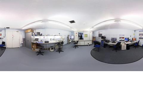
Virtual Tour - Scanning room LA62
360° Virtual Tour of University of Southampton X-ray Histology Facility - Scanning room LA62
- Setting the foundations for 3D X-ray histology (XRH) -
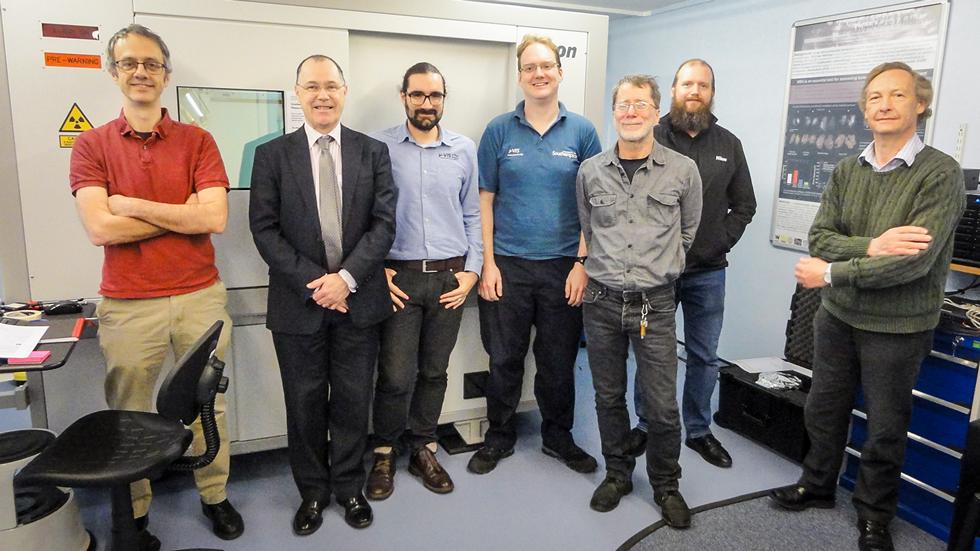
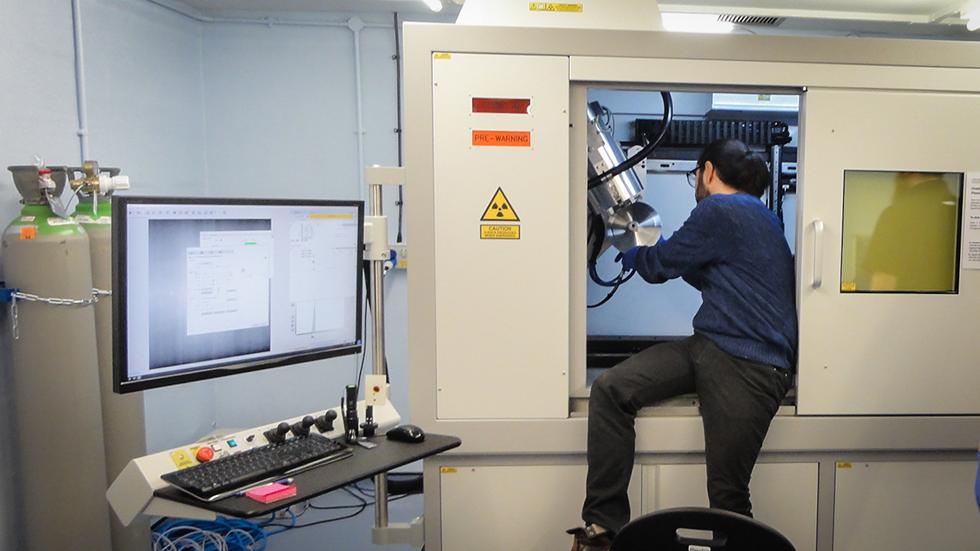
This is a custom-designed µCT scanner optimised for high throughput 3D X-ray Histology. This scanner was designed by the μ-VIS and the XRH team and built by Nikon X-Tek systems Ltd. It is the cornerstone of the second phase of development of 3D X-ray Histology, which is fully funded by Wellcome Trust (2019-2022; £1.14M; Wellcome Trust, Biomedical Resource and Technology Development Grant).
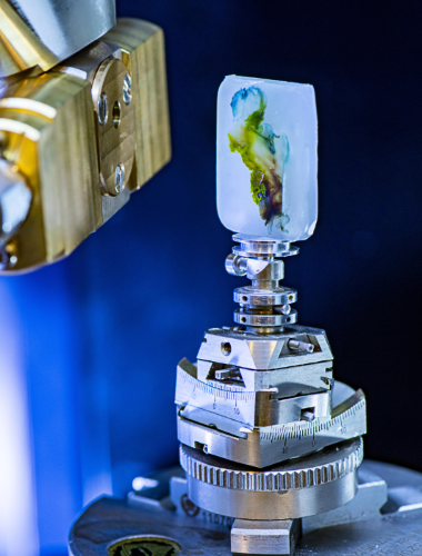
The XRH scanner can be used for non-destructive radiographical and tomographical inspection of biomedically relevant specimens, and is optimised for high-throughput imaging of unstained formalin-fixed and paraffin-embedded (FFPE) soft tissue specimens.
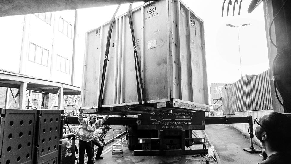
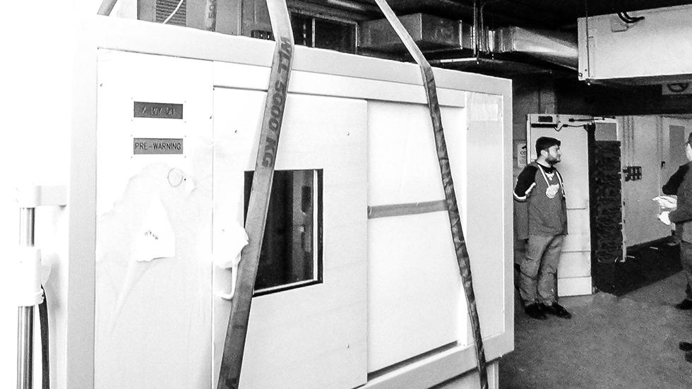
The equipment was commissioned in January, 2020 and is hosted at the Biomedical Imaging Unit at University Hospital Southampton.
‣ Nikon multi-target micro focus X-ray source
↪ 130 kVp / 130 W Mo rotating target (custom)
10 μm spot up to 50 W
↪ 180 kVp / 20 W Mo transmission targets (custom)
1.5 μm spot up to 5 W
↪ 225 kVp / 225 W multimetal reflection target
2.5 μm spot up to 7 W
‣ sample autoloader | up to 10 specimens per rack (custom)
‣ optimised for high throughput 3D X-ray Histology
‣ 2.8 x 2.8 k 16-bit flat panel detector
‣ Standard cone-beam & helical scanning
‣ samples up to ~400 mm and up to 50 kg
360° Virtual Tour of University of Southampton X-ray Histology Facility - Scanning room LA62
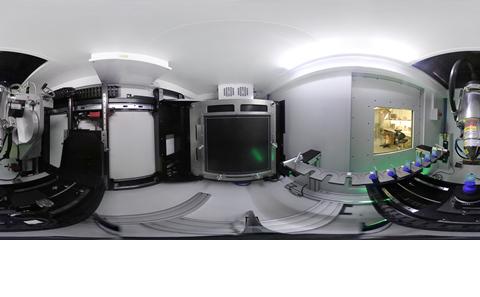
360° view inside the XRH scanner cabinet.