Stephanie K. Robinson1,2; Jonathan Ramsden2; Jane Warner2; Peter M. Lackie2; Tiina Roose1.
1 Bioengineering Sciences Research Group, Faculty of Engineering and the Environment, University of Southampton
2 Clinical and Experimental Sciences, Faculty of Medicine, University of Southampton
Overview
Lung lymphatics maintain fluid homoeostasis by providing a drainage system that returns fluid, cells and metabolites to the circulatory system. The 3D structure of the human pulmonary lymphatic network is essential to the lung function, but it is poorly characterised. Image-based 3D mathematical modelling of pulmonary lymphatic microfluidics has been limited by the lack of the accurate and representative image geometries. This is due to the microstructural similarity of the lymphatics to the blood vessel network, the lack of lymphatic-specific biomarkers, and the technical limitations associated with image resolution in 3D techniques and sectioning artefacts present in 2D techniques.
We present a method that combines lymphatic specific (D240 antibody) immunohistochemistry (IHC), optimised high-resolution X-ray microfocus computed tomography (μCT), and finite-element mathematical modelling to assess the function of human peripheral lung tissue.
These initial results identify lymphatic heterogeneity within healthy lung tissue. Significant inverse correlation exists between both lymphatic vessel volume fraction and their fractal dimension (sinuosity) with the increasing distance from the lung pleural surface (p<0.001 ,n=25 and p<0.01, n=20 respectively). Microfluidic modelling was successful, showing in healthy tissue that fluid derived from the blood vessels drain through the interstitium into the lymphatic vessel network.
This proof of principle study establishes a new workflow for further studies in the area of lung lymphatics and demonstrates for the first time the combination of correlative μCT and IHC to enable 3D mathematical modelling of human lung microfluidics at micrometre resolution.
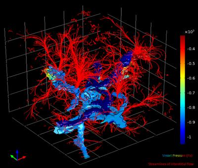
Figure 1 - 3D model simulation result for an exemplary section of lung tissue : A graphical representation of the solution for the static pressure (Pa) within the lymphatic vessels and blood vessels with healthy lung tissue. Streamlines of the Darcy’s velocity field into the interstitium are shown in red. The interstitial pressure results have been removed from view so the streamlines could be visualised.
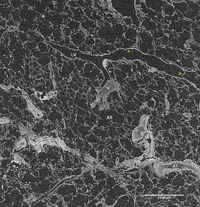
Figure 2 - Representative orthoslice of human lung tissue scanned at 6 μm pixel resolution (using Med-X prototype); LV=Precollecting lymphatic vessel, yellow arrow head= lymphatic valves, AT occluded artery, V=occluded vein, asterisk= patent blood vessels, AW= airway, AS alveolar sac