Aravinthan Vijayakumar1, Ali Mosayyebi2, Orestis Katsamenis3, Dario Carugo4
- IP undergraduate student, bioengineering group, University of Southampton
- PhD student, bioengineering group, University of Southampton
- Research Fellow, µ-VIS, University of Southampton
- Frontier Fellow, Bioengineering group, University of Southampton
The study was carried out as a part of a bigger project towards understanding the mechanism of particle deposition in stented ureter models, which was led by Dr Dario Carugo who is the leader of MiNaTher (Micro and Nano Therapies). CT imaging was conducted in the Custom 450/225 kVp Hutch at μ-VIS on a Universa® Firm (Cook® Medical, Ireland) ureteral stent. This method was preferred to obtain the geometry, as the stent geometry is quite complicated to model without any approximation. In addition to this, the stent had natural curves that would be difficult to replicate manually. Therefore, this procedure allowed a geometry that is directly comparable to the experiment. The image files that were obtained from the μ-CT scan were converted to VOL files of 32bit. These were then image processed to generate and export the stent geometry.
The image processing software that were utilised for this purpose were Fiji ImageJ and Simpleware ScanIP (Synopsys®, USA). ImageJ is an open-source image-processing package. ScanIP is an image visualisation and processing software used for volume (e.g. μ-CT, MRI) data. It is specifically known for its powerful image visualisation, analysis, segmentation and quantification tools.
ImageJ was primarily used to concatenate four raster-scans conducted consequently with ~15% overlap to increase the imaged volume. This process allowed us to image the whole stent, which could then be further processed to collect useful data.
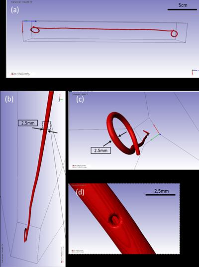
Figure: ScanIP images of the active mask (a) complete horizontal view, (b) view from above, (c) view of the pigtail, and (d) zoomed-in view of the side-hole