Archaeologists and engineers from the University of Southampton worked together with the British Museum to examine buried Roman coins using the latest X-ray imaging technology
Originally designed for the analysis of substantial engineering parts, such as jet turbine blades, the powerful scanning equipment at Southampton’s µ-VIS Centre for Computed Tomography is being used to examine Roman coins buried in three archaeological artefacts from three UK hoards.
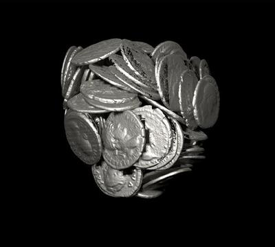
The centre’s equipment can scan inside objects – rotating 360 degrees whilst taking thousands of 2D images, which are then used to build detailed 3D images. In the case of the coins, the exceptionally high energy/high resolution combination of the Southampton facilities allows them to be examined in intricate detail without the need for physical excavation or cleaning. For those recently scanned at Southampton, it has been possible to use 3D computer visualisation capabilities to read inscriptions and identify depictions of emperors on the faces of the coins – for example on some, the heads of Claudius II and Tetricus I have been revealed.
University of Southampton archaeologist, Dr Graeme Earl says, “Excavating and cleaning just a single coin can take hours or even days, but this technology gives us the opportunity to examine and identify them quickly and without the need for conservation treatment at this stage. It also has potential for examining many other archaeological objects.
“The University’s Archaeological Computing Research Group can then take this one step further – producing accurate, high resolution CGI visualisations based on scan data. This gives archaeologists and conservators around the world the opportunity to virtually examine, excavate and ‘clean’ objects.”
Dr Roger Bland, Head of Portable Antiquities and Treasure at the British Museum comments, “This scanning technique is already yielding some fascinating results and the possibility of identifying a hoard of coins in a pot, without removing them, is very exciting. Working with archaeologists and engineers at Southampton, it is exciting to be pioneering and exploring the potential of a process which is faster, cheaper and less interventive than excavation.”
The three objects examined at Southampton are:
- A cremation urn containing nine coins, dating from AD282, found in the Cotswolds. This item in particular would take months to excavate – with archaeologists needing to carefully examine bone fragments and remains to extract more information about its past.
- An estimated 30,000 Roman coins discovered in Bath, dating to around AD270 and concreted together in a large block weighing over 100 kilograms (radiograph image only).
- A small pot dating to the 2nd century found in the Selby area of East Riding in Yorkshire.
Director of the University’s µ-VIS Centre for Computed Tomography, Professor Ian Sinclair says, “Our centre examines a wide variety of objects from the layup of individual carbon fibres in aircraft wing components, to the delicate roots of growing plants, and now ancient Roman coins. It is our integration of state-of-the-art imaging hardware, world-class computing and image processing expertise, which allows us to break new ground. We have recently formed an inter-disciplinary research group for Computationally Intensive Imaging, which brings together a broad spectrum of world-class imaging activities from disciplines across the University – of which this project is an excellent example.”
The University of Southampton and the owners of the artefacts have plans to share the scan data with the public, hopefully through future exhibitions and online.
10 July 2012