Developing 3D X-ray histology has been an exiting journey. Here we capture some of the highlights and we briefly touch on what the future might hold.
Prof. Peter JS Smith
This research is an excellent example of how funding of a small pilot project lays the foundation resulting several years later in major grant funding. This is a fantastic collaborative achievement.
Professor Peter JS Smith - Director of the Institute for Life Sciences (IfLS)
First steps
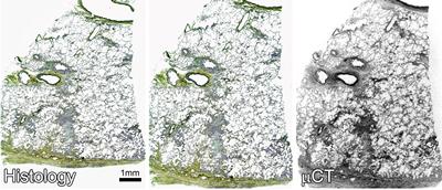
Image matching of μCT and histological section stained with Movat’s Pentachrome [Scott et al., 2015]
3D X-ray Histology (XRH) is based on the proof-of-principle study we published in 2015 [Scott et al.] initiated by Katherine Seal, a medical student and Dr Anna Scott funded by an IfLS Interdisciplinary ‘Bridging the Gap’ scheme. XRH integrates the results of more than five-years continuous development of hardware, imaging/visualisation protocols and workflow.This provides 3D visualisation compatible with both the current clinical histology workflow and archived samples.
First research applications
In 2016, samples from patients with idiopathic lung fibrosis were used to characterize the shape and interrelationships of areas of fibrosis in 3D, demonstrating the value of non-destructive 3D (volume) imaging by micro-CT. Working with the team, Dr Mark Jones, Prof Luca Richeldi and colleagues identified fibrotic structures with large variations in shape and volume, suggesting previously unrecognized plasticity that could not be identified using current ‘gold standard’ 2D histology (Jones et al., 2016; also see video below).
Simultaneously, work with Dr Tillie Hackett from the James Hogg Research Centre (University of British Columbia) provided important insights into the pathogenesis of chronic obstructive pulmonary disease (COPD) published in The Lancet Respiratory Medicine (Koo et al, 2018). This demonstrated:
‣ the ability to image standard formalin-fixed paraffin embedded tissue samples, allows access to large banks of archived diagnostic tissue ‣ volumetric micro-CT imaging helps understanding disease pathology ‣ micro-CT guided histological sectioning aids correlative imaging
Three-dimensional characterization of fibroblast foci in idiopathic pulmonary fibrosis - Jones, et al.JCI insight 1.5 (2016) - This study showed that, when seen in 3D, fibroblast foci form a constellation of heterogeneous structures with large variations in shape and volume, suggesting previously unrecognized plasticity. It also demonstrated that the application of 3D imaging methodologies can further advance concepts of disease pathogenesis.
Nikon Med-X (prototype) - a soft tissue X-ray histology prototype
Med-X (prototype)
The first studies listed above were conducted using engineering scanners and a unique highly versatile heavy-duty walk-in experimental μCT chamber, equipped with multiple micro-focus X-ray sources and detectors.
These systems provided the flexibility needed to optimise imaging conditions, and establish the proof-of-principle. However, the sheer size, cost and designs specifications of such systems are would render the technique unrealistic as a histology tool for research or clinical diagnistics.
The Med-X prototype μCT system was developed as part of a Wellcome Trust Pathfinder* project in close collaboration with Nikon X-Tek Systems Ltd. In essence Med-X is a ‘distilled’ version of its engineering ‘ancestor’, specifically designed and further optimised for use with soft tissue in a medical research/clinical environment.
Proof of utility and a robust and detailed workflow was demonstrated in our recent paper [Katsamenis et al, Am. J. Pathol] specifically aligned with the use of routinely prepared paraffin embedded tissue samples.
* Wellcome Trust/Pathfinder, 2016 -2017, Development of micro-computed tomography (μCT) for enhanced diagnosis and prognosis in interstitial lung diseases (ILD)
Foundations for routine 3D X-ray histology
The utility of 3D X-ray histology for respiratory research demonstrated in our Wellcome Trust Pathfinder project (see publications page) will be extended by a Wellcome Trust Technology Development Grant-funded project: “Foundations for routine 3D X-ray histology”.
This will develop hardware, software and workflows to:
‣ extend the range of tissues and sample types that can be scanned; ‣ significantly increase throughput, reducing scan times and cost per scan; ‣ standardise image acquisition and processing workflows; and ‣ raise awareness and drive uptake of 3D X-ray histology (XRH)
Advantages of XRH
Micrometer resolution 3D: Visualise and quantify microstructure soft tissue in their 3D context.
Non-destructive: Can be combined with specific 2D histology techniques, including histochemistry, immunocytochemistry, in situ hybridization
Volume imaging of tissue heterogeneity; Improve understanding of disease initiation and progression in 3D.
Histomics: Help identify new microstructural hallmarks of disease.
Formalin fixed paraffin embedded samples: Imaging archival material stored in many hospitals and tissue banks will allowing validation of microstructural hallmarks of disease
Fully digital 3D: XRH coupled with artificial intelligence/computer-aided diagnosis, could improve diagnostic accuracy and support patient stratification.
Video 1. Simultaneous visualization of a single μCT and hematoxylin and eosin histology slice. Coregistration of histology with μCT image data and simultaneous visualization of both data sets allow for direct comparison of the two imaging modalities and precise, histology-guided identification of a wide range of tissue structures and diagnostically relevant histologic criteria | [Katsamenis et al, Am. J. Pathol]
Video 2: Orthogonal plane view and arbitrary virtual slicing. Orthogonal planes of the μCT data can also be viewed immediately after CT reconstruction for analyzing the spatial arrangements of tissue (micro)structures, their orientation, as well as heterogeneity and connectivity in (3D) space. An example of such an interactive assessment of 3D data is shown herein, where the reviewer locates a blood vessel and examines the cross-sectional views along two orthogonal planes. | [Katsamenis et al, Am. J. Pathol]