Integrating the 3-dimensional bioelectrospray cell culture model with a microfluidic platform to model real-time physiological changes
NAMRIP's workshops put Paul Elkington in touch with Xunli Zhang, and then NAMRIP funded their collaboration, leading to the integration of Paul’s 3-dimensional bioelectrospray cell culture model into Xunli’s microfluidic platform to model real-time physiological changes.
The NAMRIP support permitted proof-of-concept experiments to take place to show that pharmacokinetic modelling can be performed in vitro in this system. The first significant output of the proof-of-concept work, a peer-reviewed publication in mBio [1], has been cited 47 times.
In keeping with NAMRIP's encouragement to translate promising research once proof-of-concept has been verified, the team started looking towards developing the technology to generate societal benefit. They have reported the translational opportunities the technology presented via the study of the effect of immune checkpoint inhibition in cancer on infection reactivation [2], and the identification of new treatment approaches [3].
This work led to the formation of new collaborations to investigate different antimicrobial compounds within the system [4].
Recently, the successful outputs of this NAMRIP project underpinned a MRC Project Grant award by the tuberculosis group entitled “Investigating local determinants of outcome in human tuberculosis MR/W025728/1 £777,561 (2022 – 2025)”. This award will fund further development of the system with single cell RNAseq approaches and real-time imaging.
In line with NAMRIP's drive to support early career researchers, the work funded by NAMRIP's pump-priming award contributed to a successful application for a University of Southampton Anniversary Fellowship for Liku Tezera, who developed the 3D microsphere model, to develop his interdisciplinary approach to identify new approaches to infectious disease.
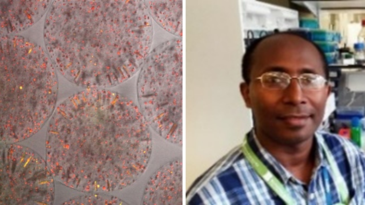
The image on the left, below, shows the microspheres with cells shown in red, and degraded collagen shown in yellow.
References
1 Bielecka, M. K. et al. A Bioengineered Three-Dimensional Cell Culture Platform Integrated with Microfluidics To Address Antimicrobial Resistance in Tuberculosis. mBio 8, e02073-02016, doi:10.1128/mBio.02073-16 (2017).
2 Tezera, L. B. et al. Anti-PD-1 immunotherapy leads to tuberculosis reactivation via dysregulation of TNF-alpha. eLife 9, doi:10.7554/eLife.52668 (2020).
3 Reichmann, M. T. et al. Integrated transcriptomic analysis of human tuberculosis granulomas and a biomimetic model identifies therapeutic targets. J Clin Invest 131, e148136, doi:10.1172/JCI148136 (2021).
4 Gonzalo, X., Bielecka, M. K., Tezera, L., Elkington, P. & Drobniewski, F. Anti-Tuberculosis Activity of Three Carbapenems, Clofazimine and Nitazoxanide Using a Novel Ex Vivo Phenotypic Drug Susceptibility Model of Human Tuberculosis. Antibiotics (Basel) 11, doi:10.3390/antibiotics11101274 (2022).