Our work on biofilms in catheters and other devices is a collabopration between the Faculties of Health Sciences, Natural and Environmental Engineering and Medicine. NAMRIP member Dr Sandra Wilks leads, along with fellow member, Professor Bill Keevil.
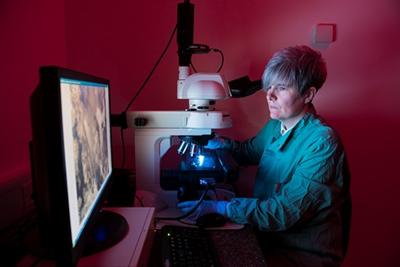
The project employs an inter-disciplinary approach and aims to increase understanding of how biofilms behave. The team examines biofilms on urinary catheters using episcopic differential interference contrast, EDIC, and microscopy imaging, and viability techniques. The study uses a combination of laboratory-based models and patient samples.
Blockages in catheters often occur from crystalline deposits formed as the pH rises due to the action of urease-producing bacteria; the most commonly found species being Proteus mirabilis. These crystalline biofilms have been found to develop on all catheter materials with P. mirabilis attaching to all surfaces and forming encrustations. This poses a high risk from urinary tract infections (UTIs) in patients, especially those with long-term indwelling catheters. Read about this work in: Sandra A. Wilks, Mandy J. Fader, C. William Keevil, Novel Insights into the Proteus mirabilis Crystalline Biofilm Using Real-Time Imaging (2015).