Imaging prize brings the natural world into clear focus
Students from across the University of Southampton have, once again, shared their biological imaging talents through an annual competition organised to honour the memory of a former academic.
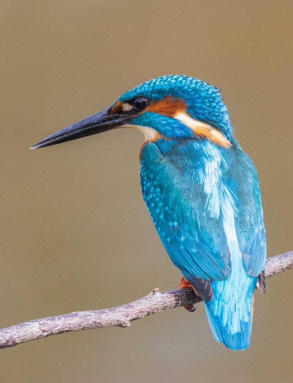
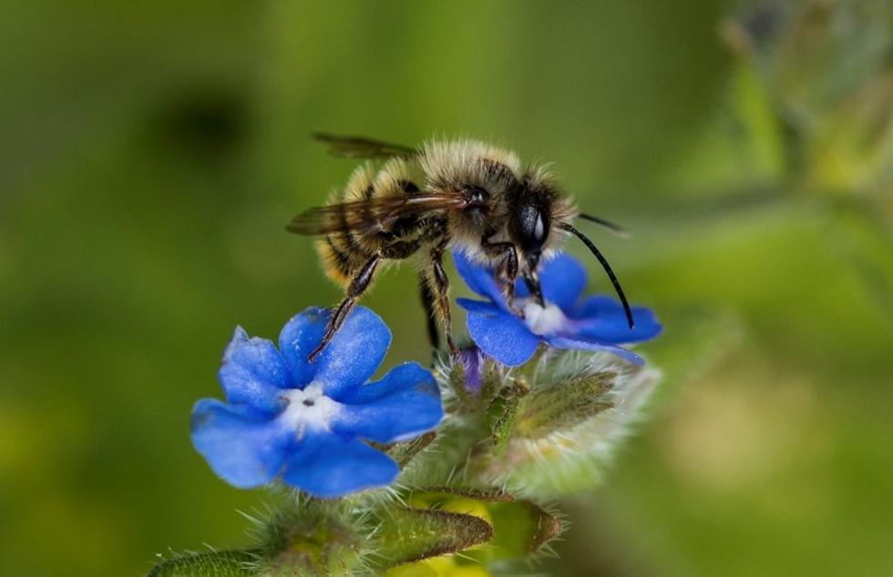
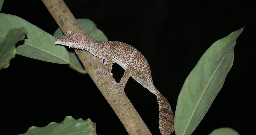
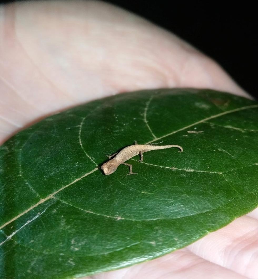
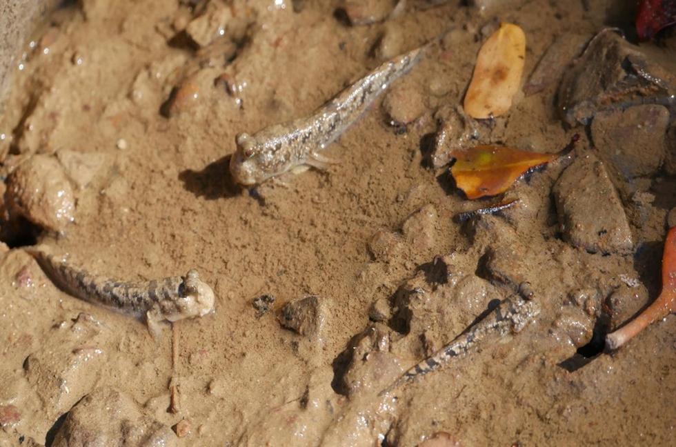
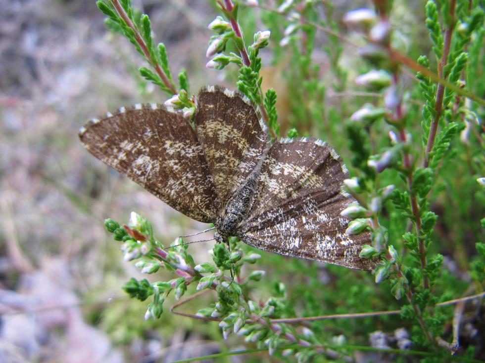
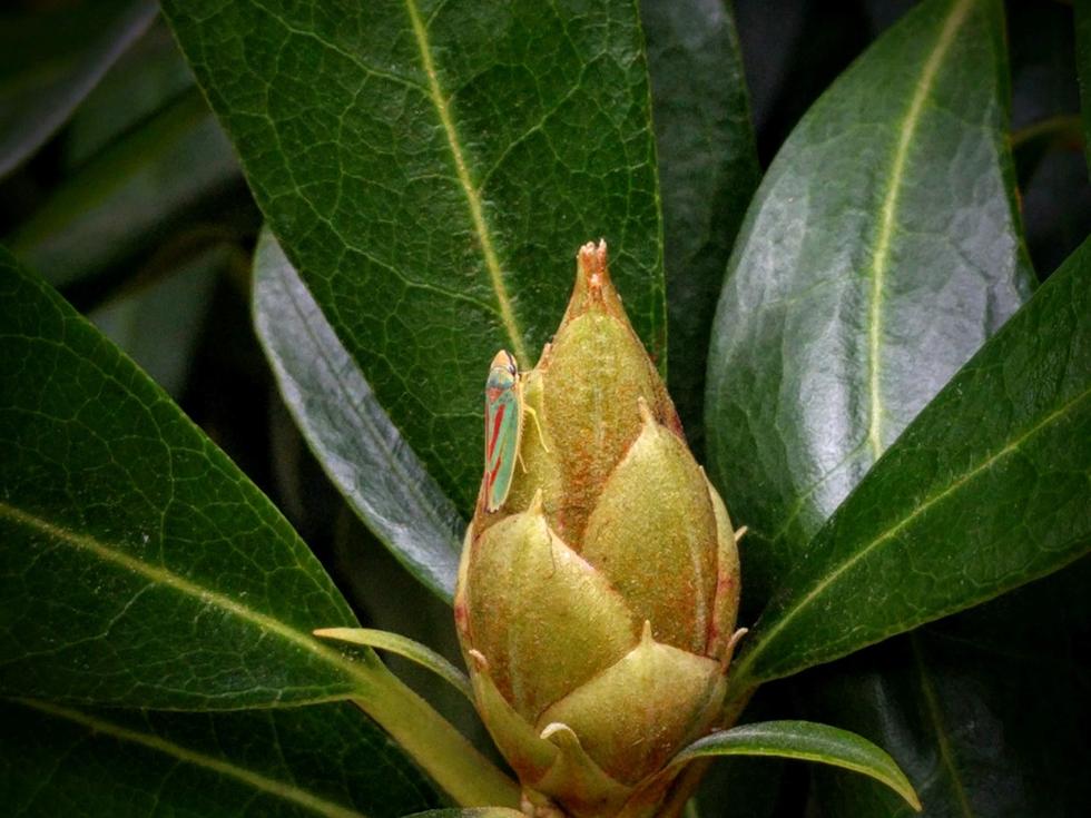
The Hans Schuppe Image Prize, managed by the University’s Imaging Microscopy Centre (IMC) is open to every undergraduate and postgraduate student in Southampton’s School of Biological Sciences to submit up to three images related to biology in the widest possible sense - from microscopic images and x-ray scans to zoological and botanical photographs of wildlife and the environment.
The Prize, established in 2016, is named in honour of Dr Hans Schuppe, founder of the state-of-the-art IMC who sadly died in 2013. Colleagues and former students remember Dr Schuppe as a deeply popular member of staff who was passionate about helping people and committed to excellence in teaching, advancing optical imaging and working with others to get the most out of their own microscopy.
The IMC’s technology supports a spectrum of scientific research – from studies investigating human health conditions to research that aims to solve environmental problems. The facility is available to resarchers across the University in areas such as biosciences, chemistry, medicine and engineering as well as external users from other universities and businesses.
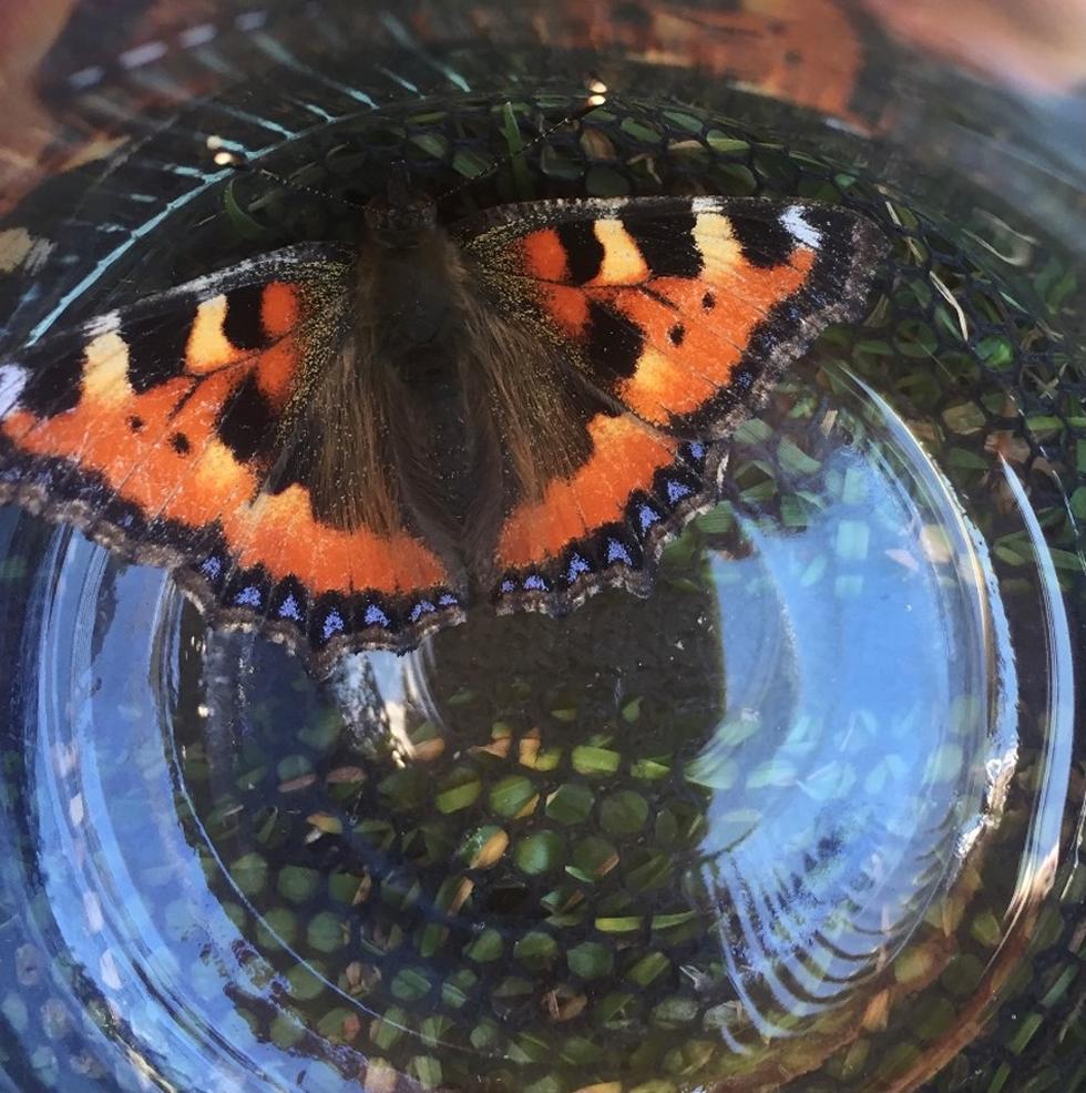
Dr Hans Schuppe Image Prize 2021
This year’s competition was judged by Dr Mark Willett, the University’s Imaging Services Manager, and Dr Neil Gostling, Lecturer in Evolution and Palaeobiology and lead for the Zoology programme.
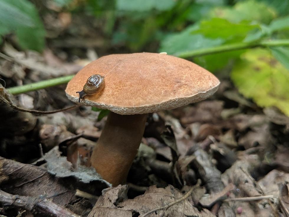
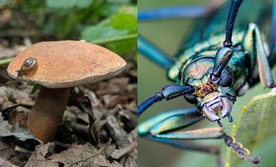
This year’s overall winner for 2021 is Magda Steele, a PhD candidate in Biological Sciences who captured a very small Bolete snail enjoying a fungal fruiting body in the woods on the University’s Highfield Campus. Although the images is not related to her research, Magda says they reflect ecology in general and show that “thriving ecocystems can be found even in the heart of the city.”
The judges loved Magda’s image which they said shows Biology in three kingdoms – fungal, animals and plants as well as the ecological interaction of all three. “To discover that it was taken on campus just shows how lucky we are with the university’s environment,” the judges commented.
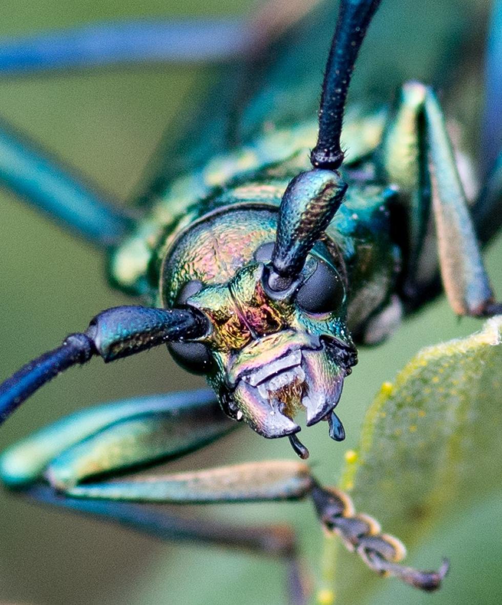
Runner-up for 2021 is Matthew Woodard, postgraduate researcher in Ocean and Earth Science, whose prize-winning image is a close-up of a Musk Beetle (Aromia mochata) found and photographed in the New Forest in 2020. Matthew says it was a real treat to find this beetle – one of the UK’s longest - on a rare day out in-between lockdowns. He adds, “they get their name from a characteristic musky smell that can be detected on leaves they have visited.”
The judges like the amazing detail of Matthew’s image which is so well focused and not only had a real sense of three-dimension but of character as well. “We don’t get to see close-ups of our native beetles like this very often and its really quite amazing to see what they actually look like,” said the judges.