The Southampton Respiratory Imaging Group has focussed particularly on developing methods for analysing aerosol deposition data with the specific aim of describing the deposition patterns in terms of the different airway generations which make up the pulmonary airway tree.
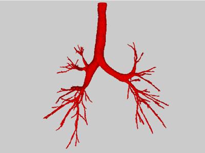
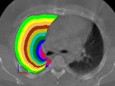
It is not possible to directly relate the airway tree anatomy to the functional imaging data that is used to measure drug deposition. Therefore, this work has led to the development of models that describe the spatial distribution of the airway tree, that can be applied to funcitonal imaging data. The most basic of these models involves dividing the lung region into a series of shells, defined by the fractional distance from the hilum. The hilum is usually chosen as the point where the main bronchous enters the lung. An example of the shells generated by the process is shown in the image here.
This work has recently expanded to include the measurement of mucociliary clearance rate. This is carried out by assessing the clearance rate from the lung of an inhaled radioactive aerosol using sequential gamma camera imaging. MCC rate is considered to be a useful biomarker in cystic fibrosis and a number of other lung diseases.