In the healthy lung, the clearance of the mucus layer created by airway secretory cells and submucosal glands occurs due to the coordinated movement of ciliated cells that act to propel the mucus out of the lung toward the mouth. In airway diseases, such as cystic fibrosis (CF), chronic bronchitis (CB) and asthma, this mechanism may be impaired, potentially leading to further exacerbation of airway inflammation.
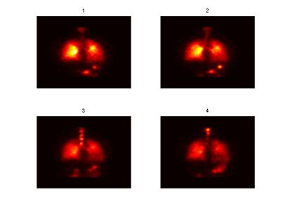
Rates of mucociliary clearance (MCC) in humans can be measured by assuming that a non-permeating, inhaled marker depositing on the airway surface will move out of the lung only as a consequence of the motion of the airway secretions. The marker can be labelled with a radio-isotope, which can then be detected using either a planar gamma camera or 3D imaging techniques such as SPECT (Single Photon Emission Computed Tomography). The image shows an example of the type of images acquired using a planar gamma camera. These images clearly show the clearance of a radio-labelled marker from the lung over the four time points (labelled 1-4). Computer analysis techniques are used to quantitatively assess the rates of clearance from this kind of data.
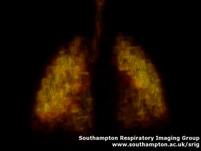
Three-dimensional imaging techniques, such as SPECT, can be used to provide a 3D analysis fo the deposition of a radio-labelled aerosol. In the same way as for the planar gammer camera images, a number of these images can be taken over a period of time and used to assess the rate of clearance from the lung. The figure shows an example 3D SPECT image.