3.5. Group I with feedback and without rule (Fr n=34)
All subjects showed a characteristic positive peak about 250msec onwards in all trial displays for both groups learners and non-learners. The positive activity peaks were greater for the learners group than the group of non-learners
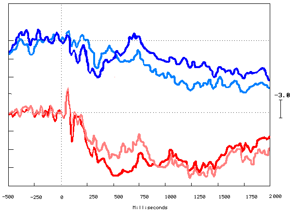
All traces are shown with reference to linked mastoids. Positivity at the named electrode denoted by downward deflection of the trace.
3.5.1. First fifty and last fifty trials:
In figure (3.5.1.) we are comparing the learners and non-learners first fifty trials out of the two hundred trials versus the last fifty trials. In the first fifty we expect they are guessing, as they have not had the chance to learn yet, and the last 50 as we can assume that they either have or have not learned.
The learners start-to-end trials comparison showed that they were different with increase in the positivity occurring from 200 msecs onwards in the last fifty displays. The last fifty traces deviated in the cognitive part from 200msecs to 900msecs. Whereas in the case of non-learners group, the activity appeared to be similar throughout the all task with insignificant differences
Figure (3.5.2.) shows that there are very clear positivity differences for the learners more than the non-learners group after learning (during the last fifty trials) started around 300msecs and ended about 1300msecs. Comparing this to the equivalent traces for the last fifty traces. The first fifty traces for both groups showed the same morphology and the learners first fifty was more positive going than the non-learners trace. This means that in the learners brain potentials for the first fifty answers provoke a more positive potential during both time windows.
Figure (3.5.1.) Grand average ERPs elicited by the learners first fifty trace in light red color and the last fifty traces in red color and the non-learners first fifty trace in light blue color and the last fifty in blue color. Recorded at FZ mid-frontal site electrode. Y-axis shows amplitude in Microvolts (
m V) and X-axis shows latency in milliseconds (ms). The black vertical dotted line marks the point of stimulus onset, and the horizontal black dotted line represents the baseline. Upward deflection represents the negativity.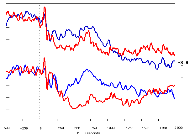
Figure (3.5.2) shows the grand averaged ERPs elicited by the learners trace in red color and the non-learners trace in blue color and the first fifty represented by the top traces and the last fifty represented by the bottom traces. Recorded at FZ mid-frontal site electrode, Y-axis shows amplitude in Microvolts (
m n ), and X-axis shows latency in milliseconds (msec). The black vertical dotted line marks the point of stimulus onset, and the horizontal black dotted line represents the baseline. Upward deflection represents the negativity.Individual electrodes gave us limited information as they reflect localized brain activity. For this reason brainmaps are useful in showing more widespread effects. Figure 3.5.3a shows the learners' top view cartoon brainmaps of made from subtraction of the first fifty trials from the last fifty trials (LFr). There were no differences recorded pre-stimulus and up to 160msec post-stimulus. The positive activity was distributed over the parietal, the temporal and occipital areas and then distributed forward to the frontal and anterior temporal areas bilaterally, and more deviated to the right-hand side. The positive activity back to decrease gradually after 500msec and localized to the left hand side posterior temporal and parietal areas and then there were no recorded differences about 900msec and beyond.
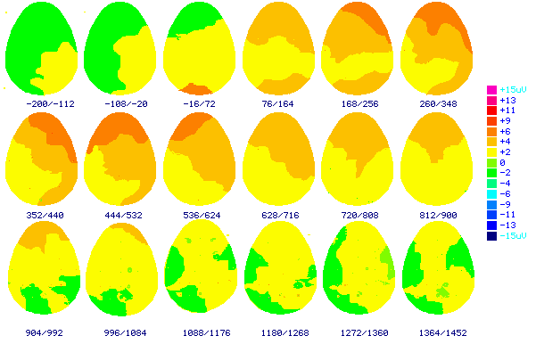
Figure (3.5.3a) shows the top view cartoon brainmaps of made from subtraction of the first fifty answers trials from the last fifty answers trials grand average ERPs elicited by the learners performance in group three (LFr). The numbers below each brainmap indicates the time in milliseconds when the map was computed. These results indicate 200msec before stimulus to 1452msec after stimulus. Color scale displays brain potentials from +15
m n in pink color, and –15 m n in dark blue. Frontal area to the top and occipital to the bottom of the brainmaps.
Figure 3.5.3b shows the non-learners top view cartoon brainmaps of made from subtraction of the first fifty trials from the last fifty trials (nLFr). There were no differences recorded pre-stimulus and up to 200msec post-stimulus. The positive activity started to appear frontally and deviated to the right-hand side. The brain activity differences did not show the same positive or negative activity distribution or latencies when compared with the learners brain maps.
Independent-Sample t-Tests for within group analysis to compare the mean amplitude for in the learners group and the non-learners group separately, and between both of them. There were high statistically significant differences between the learners start to end trials during the two time windows; the first time window learners first fifty mean amplitude was (3.02
m n ) and for last fifty was (5.3m n ); the second time window learners first fifty mean amplitude was (3.17m n ) and for the non-learners was (5.63m n ); (Table 3.5.1.), but there were no statistically significant difference for the non-learners during the both time windows (Table 3.5.1.)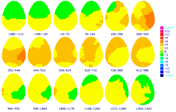
Figure (3.5.3b) shows the top view cartoon brainmaps of made from subtraction of the first fifty answers trials from the last fifty answers trials grand average ERPs elicited by the non-learners performance in group three (nLFr). The numbers below each brainmap indicates the time in milliseconds when the map was computed. These results indicate 200msec before stimulus to 1452msec after stimulus. Color scale displays brain potentials from +15
m n in pink color, and –15 m n in dark blue. Frontal area to the top and occipital to the bottom of the brainmaps.Comparing the learners and the non-learners mean amplitude during the first quarter (first fifty trials) there were no statistically significant differences (Table 3.5.2.), but after learning (last fifty trials) there was highly statistically significant difference (Table 3.5.2.).
Paired t-Tests were used for, subjects within the same group analysis to compare between the active individual electrodes. Table (3.5.3.) shows that there were statistical significant differences results in the learning group during the two time windows the last fifty answer trials versus the first fifty answer trials for these two frontal electrodes F3 & F4, and the frontal midline electrode FZ. The non-learning the last fifty versus the first fifty answer trials did not show any statistical significant differences for any of the compared electrode site during the two time windows.
|
Group |
Learners (LFr n=18) |
Non-learners (nLFr n=16) |
||
|
Time window |
1st window |
2nd window |
1st window |
2nd window |
|
First fifty (Amplitude m n ) |
3.02 |
3.17 |
3.50 |
3.30 |
|
Last fifty (Amplitude m n ) |
5.30 *** |
5.63*** |
3.70 |
3.81 |
Table (3.5.1.) Independent T-test results of the first fifty versus the last fifty answer trials mean amplitude during the both time windows for the Learners and the non-learners. *
£ 0.05, ** £ 0.01, *** £ 0.001.
|
Experiment condition |
First fifty (Amplitude m n ) |
Last fifty (Amplitude m n ) |
||
|
Group / windows |
1st window |
2nd window |
1st window |
2nd window |
|
Non-learners (nLFr n=16) |
3.50 |
3.30 |
3.70 |
3.81 |
|
Learners (LFr n=18) |
3.02 |
3.17 |
5.30 ** |
5.63 ** |
Table (3.5.2.) Independent T-test results of the learners versus the non-learners mean amplitude during the both time windows for first fifty and the last fifty answer trials. *
£ 0.05, ** £ 0.01, ***£ 0.001.|
Group |
Learners (LFr n=18) |
Non-learners (nLFr n=16) |
||
|
Electrodes sites |
1st window t taild value
|
2nd window t taild value |
1st window t taild value |
2nd window t taild value |
|
C3 |
0.59 |
1.2 |
1.2 |
0.89 |
|
C4 |
1.2 |
1.4 |
1.3 |
0.53 |
|
CZ |
1.23 |
1.3 |
2.1 |
0.54 |
|
FCZ |
1.88 |
1.77 |
1.98 |
0.97 |
|
F3 |
3.30* |
3.54 ** |
2.1 |
0.48 |
|
F4 |
3.8* |
3.60 * |
2.3 |
0.52 |
|
F7 |
1.17 |
1.11 |
1.8 |
1.4 |
|
F8 |
1.57 |
1.13 |
1.7 |
0.74 |
|
FZ |
3.89** |
3.94 ** |
2.3 |
2.4 |
|
P3 |
2.3 |
2.3 |
0.90 |
0.64 |
|
P4 |
2.5 |
2.1 |
1.1 |
0.98 |
|
TPL |
1.26 |
1.3 |
1.1 |
2.20 |
|
TPR |
1.65 |
1.80 |
1.3 |
2.3 |
|
PZ |
1.12 |
1.46 |
0.98 |
0.58 |
|
POZ |
1.11 |
1.56 |
0.99 |
0.87 |
|
T3 |
1.25 |
1.30 |
1.3 |
1.5 |
|
T4 |
1.03 |
1.5 |
1.2 |
1.6 |
|
T5 |
1.15 |
1.7 |
1.7 |
1.20 |
|
T6 |
1.12 |
1.5 |
2.1 |
1.9 |
|
ATL |
1.07 |
1.12 |
0.89 |
0.90 |
|
ATR |
1.61 |
1.8 |
1.1 |
1.30 |
Table (3.5.3.) shows Paired T-test results of the start versus the end answers trials at the individual electrode sites during both time windows for the learners and the non-Learners. *
£ 0.05, ** £ 0.01, *** £ 0.001.3.5.2.Correct versus incorrect trial answers:
The learners ERPs correct answer trials versus the incorrect answer trials seem to be different with a very clear increase in the positivity for the correct answers. These cognitive deviations start at 200msecs and end about 1200 msec (Figure 3.5.4.). Table (3.5.4.) shows that these mean amplitude differences are highly statistically significant during both time windows.
For the non-learning group both waves for the correct and incorrect answers look similar to each other (Figure 3.5.4.), but there was a minimal increase in the positivity for the correct answers starting at 400msec and ending around 700 msec. These differences are shown in the Table (3.5.4.) and these are not statistically significant during time window.
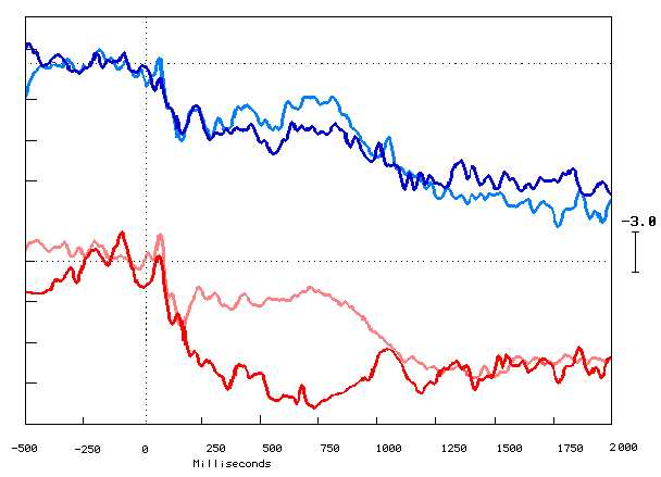
Figure (3.5.4.) the grand averaged ERPs elicited by the learners' correct answers trace in red color and the incorrect answer trace in light red color represented by the bottom traces and the non-learners correct answers trace in blue color and the incorrect answers in light blue color. Recorded at FZ mid-frontal site electrode, Y-axis shows amplitude in Microvolts (
m n ), and X-axis shows latency in milliseconds (msec). The black vertical dotted line marks the point of stimulus onset, and the horizontal black dotted line represents the baseline. Upward deflection represents the negativity
|
Group |
Learners (LFr n=18) |
Non-learners (nLFr n=16) |
||
|
Experiment condition |
1st window |
2nd window |
1st window |
2nd window |
|
Incorrect (Amplitude m n ) |
2.53 |
2.32 |
2.26 |
1.14 |
|
Correct (Amplitude m n ) |
5.55 ** |
5.70 ** |
2.38 |
2.70 |
Table (3.5.4.) Independent T-test results for incorrect versus correct answers during both time windows for the learners and non-learners group. *
³ 0.05, ** ³ 0.01, *** ³ 0.001.Comparing the correct answer trials trace of both groups, the learners group starts to show positively about 200msec and that deviation ends by 1100msec
(Figure 3.5.5.),Table (3.5.5.) shows that there are mean amplitude statistical significant differences between both groups, and that statistical differences were highly significant during the two time windows. The incorrect answer trial traces comparison between both learners and non-learners (Figure 3.5.5.) shows that the wave morphology looked very similar throughout the trial. Table (3.5.5.) confirms that there were no statistical significant differences between the mean amplitudes during the two time windows.
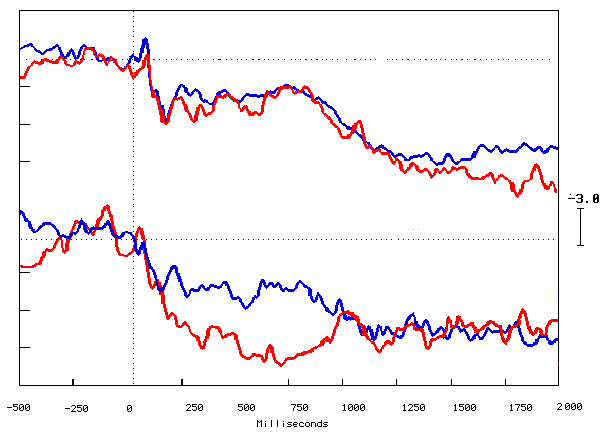
Figure (3.5.5.) Grand average ERPs elicited by the learners' traces in red color and the non-learners trace in blue color and the incorrect answer trials represented by the top traces and the correct answer trial represented by the bottom traces. Recorded at FZ mid-frontal site electrode, Y-axis shows amplitude in Microvolts (
m n ), and X-axis shows latency in milliseconds (msec). The black vertical dotted line marks the point of stimulus onset, and the horizontal black dotted line represents the baseline. Upward deflection represents the negativity.
|
Experiment condition |
Incorrect (Amplitude m n ) |
Correct (Amplitude m n ) |
||
|
Group / windows |
1st window |
2nd window |
1st window |
2nd window |
|
Non-learners |
2.26 |
1.14 |
2.38 |
2.70 |
|
Learners |
2.53 |
2.32 |
5.55 *** |
5.70 *** |
Table (3.5.5.) Independent T-test results of the learners versus the non-learners group in the incorrect and
correct answers mean amplitudes during the first and second times windows.*
£ 0.05, ** £ 0.01, *** £ 0.001.Figure (3.5.6.) shows the cartoon brainmaps of the correct answer trials concludes that there were no significant brain activities over all the brain areas from 500msec pre-stimulus. The post-stimulus the positive activity appeared over the occipital and parietal areas bilaterally. The positive activities extended forward from the right-hand side of the brainmaps over the frontal, central and anterior temporal areas from about 200msec and the positivity increased gradually over the frontal area especially to the right-hand side. The posterior temporal and occipital areas bilateral negative activities appeared from about 300msec up to 800msec post-stimulus. The positive and negative activities back to decrease gradually to the end of the task. The brain activity was greater for the correct answer trials than the incorrect answer trials.
Figure (3.5.7.) shows the cartoon brainmaps of the incorrect answers trials where there was no remarkable significant positive or negative brain activity 500msec pre-stimulus. Up to 200msec post-stimulus there was negative activity located frontally followed by positive activity bilaterally and positive activity over the occipital and parietal area bilaterally. The positive activity then distributed forward centrally and to the right. The significant positive activity very obvious over the frontal area from about 200msec to the right-hand side and then distributed bilaterally and to the anterior temporal area. The negative activity localized to the right-hand side over the frontal and the temporal areas from about 1000msec and beyond onward. Generally the brain significant activity started to decrease gradually from 1100msec to the end of the task. The overall brain activity was less than the brain activity for the correct answer trial brainmaps. Comparison with the learners correct answers cartoon brainmaps in figure (3.5.6) shows that there is a stronger and more frontal positivity.
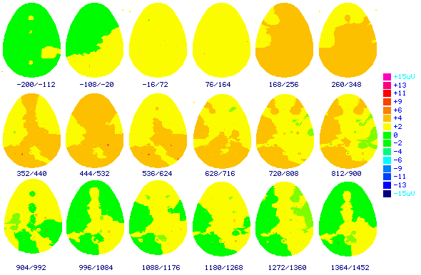
Figure (3.5.6.) shows the top view cartoon brainmaps made from the grand average ERPs elicited by the learners correct answer trials. The numbers below each brainmap indicates the time in milliseconds when the map was computed. These results indicate 200msec before stimulus to 1452msec after stimulus. Color scale displays brain potentials from +15
m n in pink color, and –15 m n in dark blue. Frontal area to the top and occipital to the bottom of the brainmaps.
Figure (3.5.7.) shows the top view cartoon brainmaps made from the grand average ERPs elicited by the learners incorrect answer trials. The numbers below each brainmap indicates the time in milliseconds when the map was computed. These results indicate 200msec before stimulus to 1452msec after stimulus. Color scale displays brain potentials from +15
m n in pink color, and –15 m n in dark blue. Frontal area to the top and occipital to the bottom of the brainmaps.Table (3.5.6.) shows that there were statistically significant differences results in the learning group during the two time windows the correct answer trials versus the incorrect answer trials mean amplitudes for these two frontal electrodes F3 & F4, one right tempro-parietal electrode TPR, and two frontal and parietal midline electrodes FZ &PZ. The non-learners for the correct versus the incorrect answer trials were not statistically significant different for any of the compared electrodes site during the two windows, except only for the frontal midline one FZ during the second time window between the correct and incorrect answers.
|
Group |
Learners (LFr n=18) |
Non-learners (nLFr n=16) |
||
|
Electrodes sites |
1st window t value |
2nd window t value |
1st window t value |
2nd window t value |
|
C3 |
0.61 |
0.44 |
1.54 |
1.3 |
|
C4 |
1.35 |
1.30 |
1.3 |
1.4 |
|
CZ |
0.23 |
1.17 |
1.22 |
1.17 |
|
FCZ |
2.03 |
1.90 |
1.34 |
1.29 |
|
F3 |
3.31 * |
3.43 * |
1.54 |
2.4 |
|
F4 |
4.82 * |
3.70 * |
1.94 |
1.2 |
|
F7 |
0.17 |
0.15 |
1.64 |
2.6 |
|
F8 |
0.61 |
0.67 |
1.26 |
2.1 |
|
FZ |
3.63 * |
3.54 * |
1.5 |
2.67 |
|
P3 |
0.38 |
0.47 |
0.5 |
1.08 |
|
P4 |
0.94 |
1.15 |
0.215 |
0.56 |
|
TPL |
1.4 |
1.70 |
1.3 |
2.2 |
|
TPR |
2.34* |
2.40 * |
1.34 |
1.92 |
|
PZ |
2.35* |
2.30 * |
1.07 |
0.34 |
|
POZ |
2.20 |
1.99 |
1.58 |
0.45 |
|
T3 |
0.57 |
0.47 |
0.65 |
1.14 |
|
T4 |
0.15 |
0.60 |
1.27 |
1.13 |
|
T5 |
0.04 |
0.42 |
1.35 |
2.40 |
|
T6 |
1.25 |
1.16 |
1.8 |
2.20 |
|
ATL |
0.34 |
0.44 |
2.05 |
1.19 |
|
ATR |
1.2 |
1.3 |
1.4 |
2.1 |
Table (3.5.6.) shows Paired T-test results of the incorrect answer versus the correct answers trials at the individual electrode sites during the both time windows for the learners and the non-Learners group.
* £ 0.05, ** £ 0.01, *** £ 0.001.3.5.3.Regional differences:
The learners' right-hand side compared with the left-hand side electrodes sites over all the brain areas. The trace morphology looks very similar 500msec pre-stimulus and 200msec post-stimulus. The positive and negative peaks started to appear about 200msec post-stimulus onward to about 1000msec with some differences from site to site (Figures 3.5.8a, b & c).
Paired t-Tests were used to compare the subjects within the same group. Table (3.5.7.) shows the comparison between the right-hand side with the left-hand side electrodes for the learners during the last fifty answer trials. There were highly statistically significant differences for the central area C4-C3, Frontal area F4-F3 & F8-F7, and Parietal area P4-P3 during the two time windows. The non-learners did not show inter-regional statistical significant differences.
|
Group |
Learners (LFr n=18) |
Non-learners (nLFr n=16) |
||
|
Electrodes sites |
1st window t value |
2nd window t value |
1st window t value |
2nd window t value |
|
C4 - C3 |
1.145 *** |
1.657 ** |
0.125 |
0.614 |
|
F4 - F3 |
0.674 ** |
1.732 ** |
0.058 |
0.155 |
|
F8 - F7 |
1.904 *** |
1.773 *** |
1.600 |
1.837 |
|
O2 - O1 |
0.506 |
0.631 |
1.000 |
0.627 |
|
P4 - P3 |
0.665** |
0.549 ** |
0.550 |
0.895 |
|
TPR - TPL |
0.021 |
0.447 |
0.445 |
0.970 |
|
T4 - T3 |
0.073 |
0.431 |
1.030 |
0.545 |
|
T6 - T5 |
0.407 |
0.792 |
1.370 |
1.223 |
|
AT - ATL |
0.343 |
0.331 |
1.631 |
1.333 |
Table 3.5.7. Paired T-test results of the right-hand side versus the left-hand side electrodes for the last fifty trials (4th quartile) during both time windows in the Learners and the non-learners group.
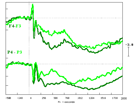
Figure (3.5.8a) shows the grand average ERPs elicited during the last fifty trials of the learning task. The right-hand site electrode site trace in dark green color and the left-hand site electrode site trace in light green color. Recorded at F4-F3, & P4-P3, refer to linked mastoids. Y-axis shows amplitude in Microvolts (
m n ), and X-axis shows latency in milliseconds (msec). The black vertical dotted line marks the point of stimulus onset, and the horizontal black dotted line represents the baseline. Upward deflection represents the negativity.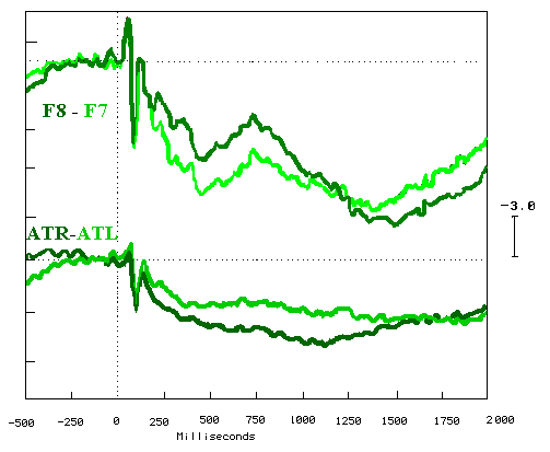
Figure (3.5.8b) shows the grand average ERPs elicited during the last fifty trials of the learning task. The right-hand site electrode site trace in dark green color and the left-hand site electrode site trace in light green color. Recorded at F8-F7, & ATR-ATL, refer to linked mastoids. Y-axis shows amplitude in Microvolts (
m n ), and X-axis shows latency in milliseconds (msec). The black vertical dotted line marks the point of stimulus onset, and the horizontal black dotted line represents the baseline. Upward deflection represents the negativity.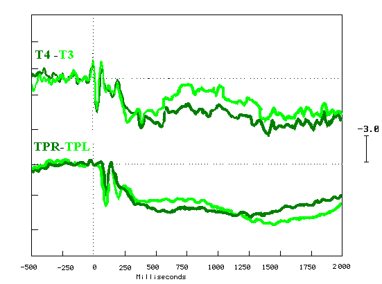
Figure (3.5.8c) shows the grand average ERPs elicited during the last fifty trials of the learning task. The right-hand site electrode site trace in dark green color and the left-hand site electrode site trace in light green color. Recorded at T4-T3, & TPR-TPL, refer to linked mastoids. Y-axis shows amplitude in Microvolts (
m n ), and X-axis shows latency in milliseconds (msec). The black vertical dotted line marks the point of stimulus onset, and the horizontal black dotted line represents the baseline. Upward deflection represents the negativity.Figures 3.5.9a & b showed the comparison between cartoon brainmap the right-hand side view versus the left-hand side view for the correct answers trials during the two time windows 250msec-550msec and 550msec-850msec. The positive activity was localised frontally and distributed backward to the anterior temporal and was more for the right-hand side view brainmaps during both time windows when compared to the left-hand side view. The activity equally distributed over the parietal, the central, the posterior temporal and the occipital areas for the correct and the incorrect answers trials and during the both windows. The positive activities were more for the second time interval window 550msec to 850msec than the first time interval window250msec to 550msec.
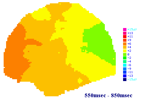
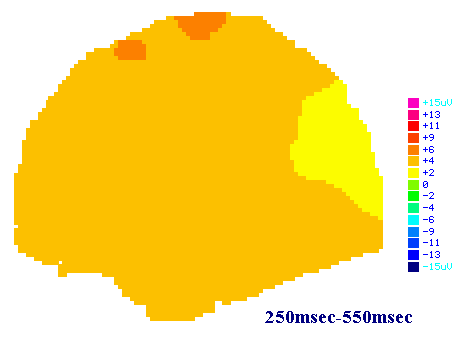
Figure (3.5.9a) shows the left-hand side view brainmaps made from the grand average ERP elicited with last fifty stimuli in the learners. The numbers below each large map indicates the time in milliseconds when the map was computed. The first time interval 250msec to 550msec and the second time interval 550msec to 850msec post-stimulus. Color scale displays brain potentials from +15 m n in pink color, and –15 m n in dark blue.
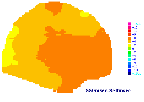
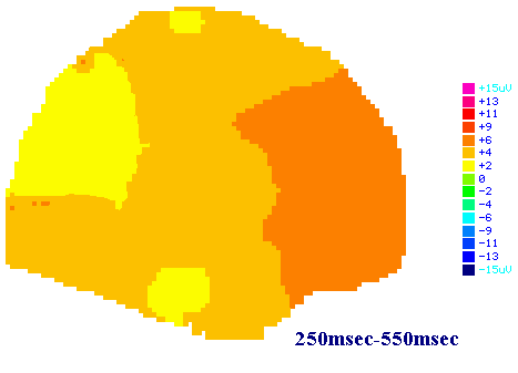
Figure (3.5.9b) shows the right-hand side view brainmaps made from the grand average ERP elicited with last fifty stimuli in the learners group. The numbers below each large map indicates the time in milliseconds when the map was computed. The first time interval 250msec to 550msec and the second time interval 550msec to 850msec post-stimulus. Color scale displays brain potentials from +15 m n in pink color, and –15 m n in dark blue.