3.6. Group II with feedback and with rule (FR n=15).
Group II subjects had been taught the differences between the patterns but had not practiced the task. All achieved the learning level quickly and there were no non-learners.
The grand average ERP showed a similar morphology to those of group I (Fr). The positivity began at 200msec and was frontally and centrally located with emphasis on the right side. Overall the positivity was of lower amplitude.
3.6.1. First fifty and last fifty trials:
The initial potentials were the same with first fifty, last fifty, correct answers, incorrect trial waveforms up to 230 msec.
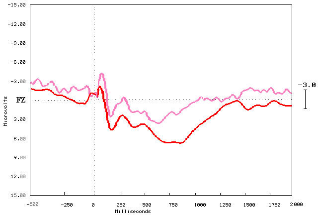
Figure (3.6.1) compares the first fifty and the last fifty trials. These subjects were already performing above 70% correct for the first fifty and achieved over 80% during the last fifty trials. The last fifty trials trace was more positive going than the fifty trial traces starting from 200msec to beyond about 1100msec.
The last fifty trials elicited a first positive peak at 220msec and another positive peak at 350msec, and the biggest one at 650msec. The first fifty elicited the same positive peaks but they were less in amplitude and earlier in latency.
The first fifty trial cartoon brainmaps subtracted from the last fifty trials showed the difference between the subjects who knew the task and when the subject knew how to perform it. There was not much difference between the first fifty and the last fifty pre-stimulus and post-stimulus till about 150msec. The positivity was more localized frontally to the right-hand side. The positive activity was distributed bilaterally over the anterior temporal areas and the frontal areas from about 200msec and extended backward over the central area. Some positivity was obvious in the posterior temporal area to the right-hand side of the brainmaps (figure 3.6.2).
Figure (3.6.1) Grand average ERPs elicited by the first fifty in light red and the last fifty in dark red. Recorded at (FZ), Y-axis shows amplitude in Microvolts (
m n ), and X-axis shows latency in milliseconds (ms). The black vertical dotted line marks the point of stimulus onset, and the horizontal black dotted line represents the baseline. Upward deflection represents the negativity.
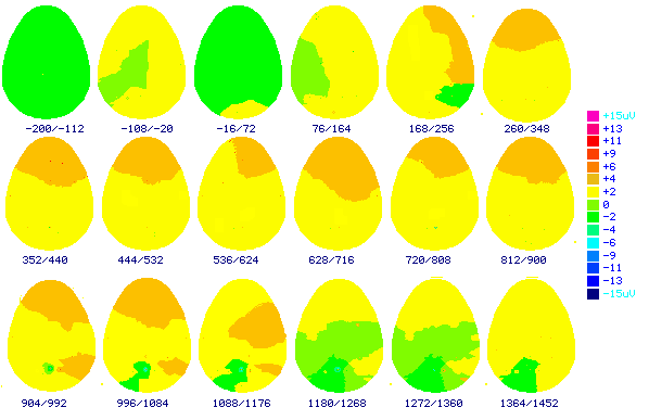
Figure (3.6.2) shows the top view cartoon brainmaps made from grand average ERP elicited by the first fifty trials subtracted from the last fifty trials stimuli. The numbers below each brainmap indicates the time in milliseconds when the map was computed. These results indicate 200 msec before stimulus to 1452 msec after stimulus. Color scale displays brain potentials from +15
m n in pink color, and –15 m n in dark blue. Frontal to the top, occipital to the bottom of the brain maps3.6.2. CORRECT VERSUS INCORRECT:
The correct answer trials were more positive going than those were to the incorrect answer trials starting from 200msec to about 1200msec.
Figure (3.6.3) shows that the incorrect and correct answers wave were very similar pre-stimulus and post-stimulus till about 250msec. The correct answers and the incorrect answers elicit positive peak at 480msec but more positive for the correct answers. The second positive peaks were about 750msec and more positive for the correct trace.
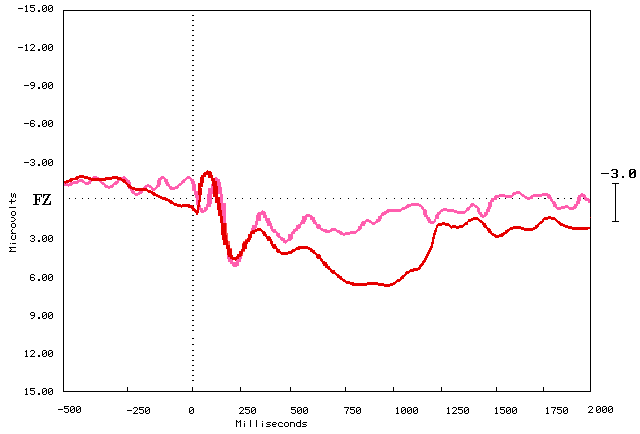
Figure (3.6.3.) Grand average ERPs elicited the correct answers in red color, and the incorrect answers in light red color. Recorded at (FZ), Y-axis shows amplitude in Microvolts (
m n ), and X-axis shows latency in milliseconds (ms). The black vertical dotted line marks the point of stimulus onset, and the horizontal black dotted line represents the baseline. Upward deflection represents the negativity.The cartoon brainmaps (Figure 3.6.4) of the incorrect answers trials showed that there was no significant positive or negative brain activity pre-stimulus. Post-stimulus there was negative activity located frontally and positive activity over the occipital and parietal area bilaterally from 200msec and then distributed forward centrally and to the right. The significant positive activity very obvious over the frontal area from about 500msec to the right-hand side and then distributed bilaterally. The negative activity bilaterally over the occipital and the parietal area. The brain activity started to decrease gradually from 1200msec to the end of the task.
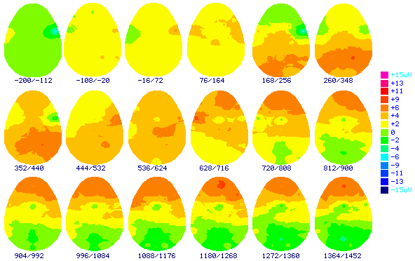
Figure (3.6.4) shows the top view cartoon brainmaps made from the grand average ERP elicited with the incorrect trials stimuli. The numbers below each brainmap indicates the time in milliseconds when the map was computed. These results indicate 200msec before stimulus to 1500msec after stimulus. Color scale displays brain potentials from +15
m n in pink color, and –15 m n in dark blue. Frontal to the top, occipital to the bottom of the brain maps.Figure 3.6.5 showed that the brainmaps of the correct answers conclude that there were no significant brain activities over all the brain areas pre-stimulus. The post-stimulus the positive activity appeared over the occipital and parietal and the negative activity over the frontal areas bilaterally. The positive activities extended forward from the right hand side of the brain maps over the frontal, central and anterior temporal areas from about 300msec and the positivity increased gradually over the frontal area especially to the right-hand side. The parietal, posterior temporal and occipital bilateral negative activities appeared from about 700msec. The positive and negative activities back to decrease gradually to the end of the task. Generally the brain positive activity was more for the correct answers trials than the incorrect answers trials.
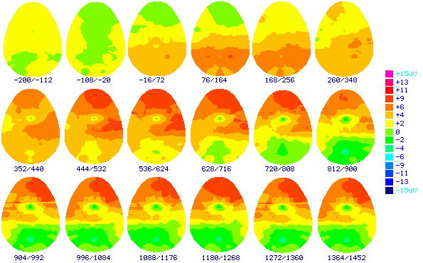
Figure (3.6.5) shows the top view cartoon brainmaps made from the grand average ERP elicited with the correct trials stimuli. The numbers below each brainmap indicates the time in milliseconds when the map was computed. These results indicate 200msec before stimulus to 1500msec after stimulus. Color scale displays brain potentials from +15
m n in pink color, and –15 m n in dark blue. Frontal to the top, occipital to the bottom of the brain maps.3.6.3. Regional differences:
Multiple equal numbers of electrodes on each side of the scalp were used to record the ERP. Comparison between ERP elicited by the correct versus the incorrect and last fifty versus the first fifty trials answers showed the differences were more prominent in the frontal right-hand side than the left-hand side. These differences begin around 250msec post-stimulus and persist beyond the recording epoch of 1000msec in the right-hand hemisphere. The central electrodes shows differences started around 300msec and end very quickly at 500msec which I considered from my inspection and comparisons with other sides electrodes not significantly different where the traces were similar throughout the task. The parietal area electrodes showed positive differences starting around 450msec to beyond 1000msec. Amplitude of P300 was maximal in frontal and higher for the correct and last fifty trial answers and P600 showed maximal amplitude for correct last fifty in the parietal locations (Figure 3.6.6).
Table (3.6.1.) shows the comparison between the right-hand side with the left-hand side electrode for the learners during the last fifty trials. There was statistically significant difference for the frontal area electrodes F4-F3 during the two time windows.
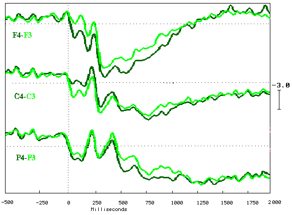
Figure (3.6.6) Grand average ERPs elicited by all subject’s performance in group II, recorded at F3-F4, C3-C4, and P3-P4 refer to linked mastoids, during the last fifty trials, Right-hand side traces represented in dark green color and left-hand side represented in light green color. Y-axis shows amplitude in Microvolts (
m n ), and X-axis shows latency in milliseconds (ms). The black vertical dotted line marks the point of stimulus onset, and the horizontal black dotted line represents the baseline. Upward deflection represents the negativity.
|
Group |
Learners (LFR n=15) |
|
|
Electrodes sites |
1st window t value |
2nd window t value |
|
C4 - C3 |
1.46 |
2.008 |
|
F4 - F3 |
2.11 * |
2.89 ** |
|
F8 - F7 |
1.66 |
1.56 |
|
O2 - O1 |
1.27 |
1.63 |
|
P4 - P3 |
1.65 |
2.01 |
|
TPR - TPL |
1.28 |
1.88 |
|
T4 - T3 |
1.78 |
0.99 |
|
T6 - T5 |
1.29 |
0.87 |
|
AT - ATL |
1.23 |
1.09 |
Table (3.6.1.) Paired T-test results of the right-hand side versus the left-hand side electrodes for the last fifty trials (4th quartile) during both time windows
Figures (3.6.8a & b) showed the comparison between the right-hand side view versus the left-hand side view for the subtraction of the incorrect answers trials from the correct answers trials brainmaps during the two time windows. The positive activity was localized frontally and distributed backward to the anterior temporal and central areas. The activity distributed over the parietal, the central, the posterior temporal and the occipital areas which means that these areas were involved in the trials performance by the same value during both conditions task correct and incorrect answers trials. These positive and negative activities were more for the first time interval window 250msec to 550msec than the second time interval window 550msec to 850msec.
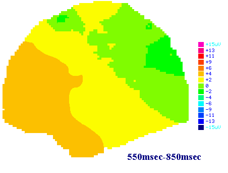
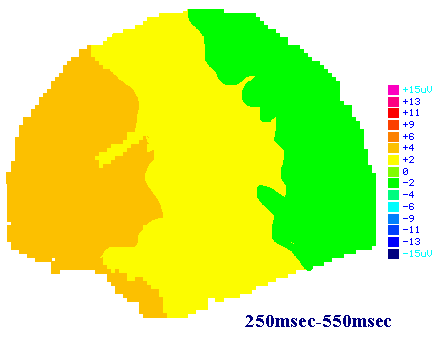
Figure (3.6.8a) shows the left-hand side view spatial brainmaps made from group two grand average subtraction ERPs elicited with the correct minus the incorrect answers trials stimuli. The numbers below each large map indicates the time in milliseconds when the map was computed. The first time interval 250msec to 550msec and the second time interval 550msec to 850msec post-stimulus. Color scale displays brain potentials from +15
m n in pink color, and –15 m n in dark blue. The frontal area to the left-hand side and occipital area to the right-hand side of the brainmap.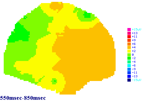
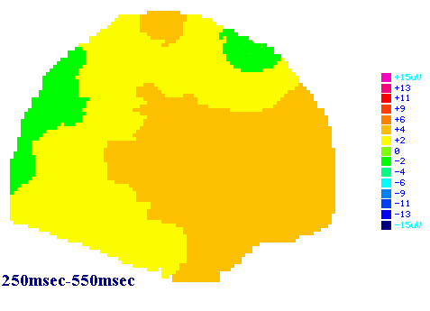
Figure (3.6.8b) shows the right-hand side view spatial brainmaps made from group two grand average subtractions ERPs elicited with the correct minus the incorrect answer trial stimuli. The numbers below each large map indicates the time in milliseconds when the map was computed. The first time interval 250msec to 550msec and the second time interval 550msec to 850msec post-stimulus. Color scale displays brain potentials from +15
m n in pink color, and –15 m n in dark blue. The frontal area to the right-hand side and occipital area to the left-hand side of the brainmap.