Clinical imaging-based 3D printing demonstrates life-saving potential in complex neurological surgery
.jpg)
A novel 3D printing workflow developed collaboratively by 3D X-ray Histology facility team at the University of Southampton and University Hospital Southampton (UHS) and has proven invaluable in a recent brain surgery case.
Southampton, Nov. 2024 – Engineers at the University of Southampton, in collaboration with clinical teams at University Hospital Southampton (UHS), have developed a novel surgical planning workflow that uses 3D-printed anatomical replicas to give surgeons a realistic, hands-on view into patient anatomy. This approach brings together advanced imaging, image processing, and 3D printing to create accurate, patient-specific anatomical models and has already shown life-saving potential in recent cases involving intracranial aneurysm surgery.
The project was led by University Hospital Southampton neurosurgeon Professor Diederik Bulters , working closely with Dr Orestis Katsamenis at the University of Southampton’s 3D X-ray Histology facility, part of the μ-VIS X-ray Imaging Centre . Together, they developed an intricately detailed 3D model of a patient’s aneurysm and surrounding bony structures. Using MRI and CT scans, the team carefully segmented and processed the 3D datasets to isolate the aneurysm and surrounding bonny structures. Professor Dennis Douroumis (University of Greenwich) team at the Centre for Research Innovation and Dr Laura Junqueira (Delta Pharmaceutics Ltd) completed the final 3D printing replicating the exact dimensions of the surgical field, using materials designed to emulate the feel of soft and hard tissues. This innovative model provided Professor Bulters’ team with an essential tactile reference that proved invaluable for preoperative planning.
According to Professor Bulters, “ We were recently faced with a particularly challenging case of a giant 7 cm intracranial aneurysm. To give reference only 3% of aneurysms are over 2.5 cm which is the conventional definition of giant. This would not have been treatable with a standard procedure using a clip to occlude the aneurysm. We hypothesised it would be possible with excision of the aneurysm and patch repair. However, given this must occur within a confined space with many geometrical constraints it was not clear if this novel approach would be technically possible. The model allowed us to simulate this and come up with the best strategy. It gave us the reassurance the procedure was technically possible. I doubt we would have had the confidence to undertake this case without being able to plan and simulate it in this way. The case went exceedingly well exactly following the plan we had made with the model, and the patient was immensely grateful and was back at home after just two nights in hospital .”
Dr Katsamenis added: “ This pioneering workflow highlights the power of collaboration between engineering and clinical teams to push the boundaries of surgical planning. Deploying our centre’s unique infrastructure and expertise we can support clinicians to set a new standard for precision in patient care. ”
As 3D imaging and printing technologies continue to advance, this approach has the potential to transform how surgeons prepare for high-risk procedures, offering patients improved outcomes and a higher level of personalized care.
Access to the μ-VIS / XRH facility was supported by the National Facility for laboratory-based X-ray computer tomography .
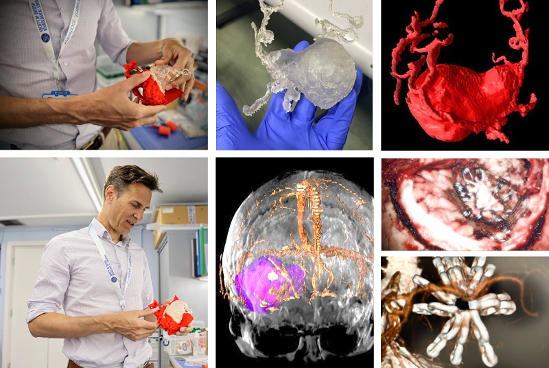
(top left, bottom left, top centre) Prof. Bulters examines the 3D-printed aneurysm and skull assembly; (top right): Digital 3D copy of the aneurysm; (bottom centre) Magnetic Resonance Angiography (MRA) 3D rendering highlighting the aneurysm (in purple) prior to surgery; (centre right, bottom left) Intraoperative image showing the aneurysm patch repair & Postoperative CT image confirming the repair)