A new study from researchers at the Centre for Cancer Immunology has discovered a new way to identify patients who are twice as likely to die from lung cancer.
The team, funded by Cancer Research UK and the Medical Research Council, used state of the art techniques to study fibroblasts, a cell that surrounds tumours. These are healthy cells that support wound healing but can be hijacked by cancer to help tumours grow and spread.
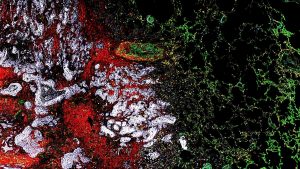
Using equipment created by engineers at the University of Southampton alongside the very latest technology, they were able to identify three different types of fibroblasts for the first time ŌĆō one ŌĆśhijackedŌĆÖ myofibroblast and two ŌĆśnormalŌĆÖ fibroblasts.
The research, published in Nature Communications, shows patients with a high proportion of myofibroblasts have double the risk of dying from lung cancer within four years compared to patients with fewer myofibroblasts in their tumours.
Dr Chris Hanley who co-led the research said: ŌĆ£These types of fibroblasts indicate whether a patient is going to survive for a longer or shorter period of time, so this information can potentially be used to give them a more accurate prognosis.
ŌĆ£We also believe that patients who have a high number of myofibroblasts in their tumour could benefit from new treatments that can target this particular cell which would ultimately improve survival rates.ŌĆØ
The team analysed 10,000 fibroblasts from 100 patients, breaking them down one by one using a technique called single cell sequencing.
They were able to capture individual cells into a droplet, before bursting the cell to capture the different molecules. This allowed them to count how many different molecules there are in a given cell, giving it a fingerprint.
This technique revealed the three types of fibroblasts, each with its own molecular fingerprint and function. These fingerprints were used to trace the fibroblastŌĆÖs origins and showed that only myofibroblasts were directly linked to increasing the risk of dying with lung cancer.
Dr Chris Hanley said: ŌĆ£What hasnŌĆÖt been clear until now is whether all fibroblasts are helpful to cancer, but this work shows that myofibroblasts are the most dangerous culprits. If we can take out myofibroblasts, we have a better chance of taking out lung cancers.ŌĆØ
This is the largest study ever conducted into fibroblasts in lung cancer patients, taking six years to complete. Dr Hanley started working with Professor Gareth Thomas on the concept long before the technological advances were in place for them to carry out their research.
Professor Thomas added: ŌĆ£We started this idea from scratch when this was a very new technology and itŌĆÖs been a long haul and an incredible amount of work to get to this point. But the insight that itŌĆÖs given us provides so many opportunities for future projects and we have a much clearer idea of where weŌĆÖre going because of this.
ŌĆ£The next step is to develop ways of targeting ŌĆśbadŌĆÖ fibroblasts to make treatments more effective in high-risk lung cancer patients.ŌĆØ
Dr Iain Foulkes, Executive Director of Research and Innovation at Cancer Research UK, said: ┬Ā ŌĆ£To beat cancer sooner, we need to understand it in detail. Thanks to pioneering research like this, we are moving closer to the day when all patients can receive treatment that is tailored to the features of their tumour. ┬Ā
ŌĆ£The technology available to cancer researchers today gives us unprecedented insights into how cancer starts and grows. We hope that this research will become the basis of new treatments for cancer in the clinic in future.ŌĆØ