Project summary
Delivery of antimicrobial drugs with nanoparticles has the potential to combat drug resistance. With conventional drug administration, the drug molecules are diluted in the body, resulting in a low concentration when they eventually reach the bacteria. But with drugs enclosed in nanoparticles, the molecules remain inside the nanoparticles until they are taken up by the bacteria.
Administration of nanoparticle-enclosed drugs has two distinct advantages. Firstly, is delivery of a larger amount of the drug to bacteria becomes possible because the drug is not diluted in the body. And secondly, nanoparticle-enclosed drugs are protected from degradation or aggregation, enabling the use of hitherto unsuitable antimicrobials.
Our Work
Nanoparticle delivery of antimicrobial drugs is a new research area, and in this project we seek to understand which type of nanoparticles can bind to bacteria. We are interested in the molecular basis of nanoparticle-bacteria interactions and we therefore use model systems that resemble the outer surface of bacteria.
Our approach is to make cell-sized liposomes, so-called giant unilamellar vesicles, of which we know the exact molecular composition. This is a well-established method, but it has not been optimized yet for glycolipids, the key molecules that define the bacterial outer membrane.
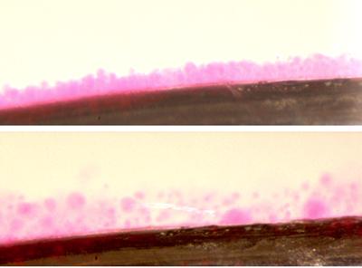
We are making these liposomes by depositing lipid molecules on platina electrodes and then applying an electric field. This electroformation protocol results in liposomes that are tens of micrometers in size. Importantly, this means that they can be easily observed with optical microscopes.
Figure 1 - Electroformation (top) and detachment (bottom) of giant liposomes with bacterial lipids.
The aim of the project is to study the binding of different types of nanoparticles to our bacteria-like liposomes, by using microscopy to visualize nanoparticle-liposome association. Because nanoparticles are synthesized from simple basic units, we can easily vary their surface characteristics to optimize liposome binding. This will enable us to understand which nanoparticle families are best suited for antimicrobial drug delivery to harmful bacteria.
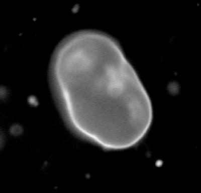
Figure 2 - Fluorescence microscopy of an individual liposome. The outer surface resembles a bacterial membrane.
About the team
Dr Maurits de Planque works in the department of Electronics and Computer Science. With a background in bionanotechnology, he works on the application of engineering methdologies to life sciences research and specializes in cell membrane research. Dr Tracey Newman is a member of the Faculty of Medicine and is based in the Centre of Biological Sciences. As a neurobiologist her research includes the development of polymersome nanoparticles to combat inflammation. Professor Robert Read is Head of the Clinical and Experimental Sciences unit of the Faculty of Medicine. His research interests include the understanding and prevention of rapidly lethal infectious diseases.