Overview
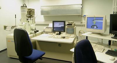
FEG SEM (Joel JSM6500)
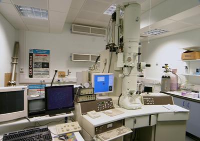
TEM (Joel JEM3010)
Electron microscopy is a technique that employs accelerated electrons to image surfaces, as the wavelength of electrons is much small than visible light (photons) the resolving power (magnification) can be much higher measurements to the nanometre scale, including nanoparticles and nanotubes.
Electron microscopes are often used to study surfaces to determine the wear and/or corrosion mechanisms that have occurred on them.
nCATS has access to two electron microscopes, one a field-emission gun scanning electron microscope (FEG-SEM) and the other a transmission electron microscope (TEM).
The Joel JSM 6500 SEM has an accelerating voltage of 0.5 to 30kV, magnifications of 10-5000,000 at a resolution of 1.5nm. It is capable of SEI and BEI imaging and EDS microanalysis. The JSM 6500 is also capable of EBSD.
JEM-3010 ultrahigh resolution analytical electron microscope with a point to point and lattice resolution of 0.17 nm and 0.10 nm, respectively.
Among its more outstanding features are a microactive goniometer with motorised axes and computer controlled data management and storage.