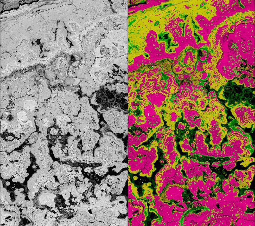
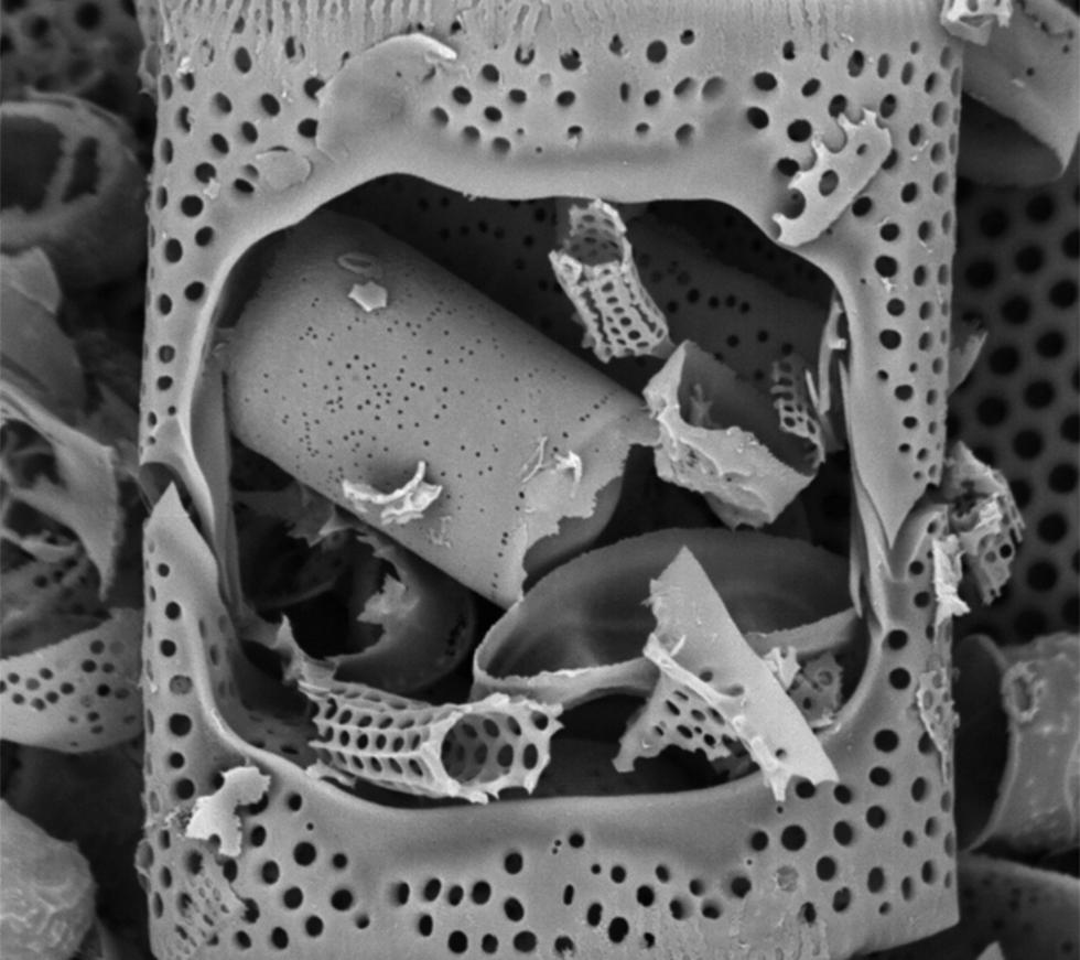
A scanning electron microscope (SEM) allows the observation of inorganic and organic materials from the nanometer- through to the micrometer-scale. It uses electrons rather than light for imaging, creating either three-dimensional-like views of a surface (secondary electron images) or images based on atomic number contrast and porosity (backscattered electron images).
The Ocean and Earth Science SEM facility is a teaching and research resource for undergraduate and postgraduate students and staff, both within Ocean and Earth Science and across the University of Southampton. More widely, it offers an imaging and analytical service, for universities and industrial organisations in the fields of geology, materials and metallurgical analysis, archaeology and environmental science. In addition to imaging, analytical services include; elemental mapping, elemental line scans and elemental SEM-EDS spot analysis (qualitative and quantitative).
Dry, non-conductive samples, for example archaeological museum pieces or beam/vacuum sensitive materials, can be both imaged and chemically analysed, without carbon or gold coating, by using the environmental SEM setup under a reduced vacuum (variable pressure mode).
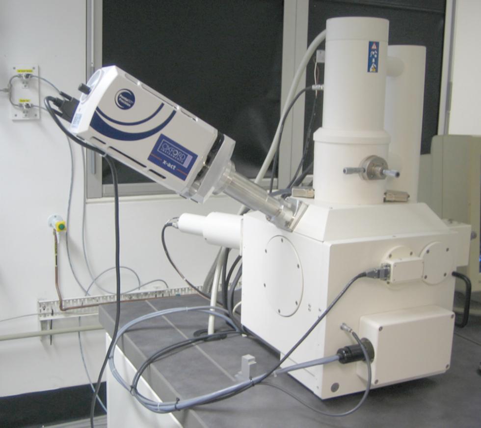
The SEM facility was established in 2001, through funding from NERC, the University of Southampton and Carl Zeiss SMT Ltd, to support high-resolution palaeoceanographical, palaeoclimatic and palaeoenvironmental research and teaching. The SEM comprises a Leo 1450VP SEM and an Oxford Instruments X-Act 10mm2 area SEM-Energy Dispersive Spectrometer (EDS), utilising the AZtec Energy software system.
To discuss your SEM requirements or for further information, contact the Lab Manager:
Dr Richard Pearce Tel: +44 (0)23 8059 6477/6518


