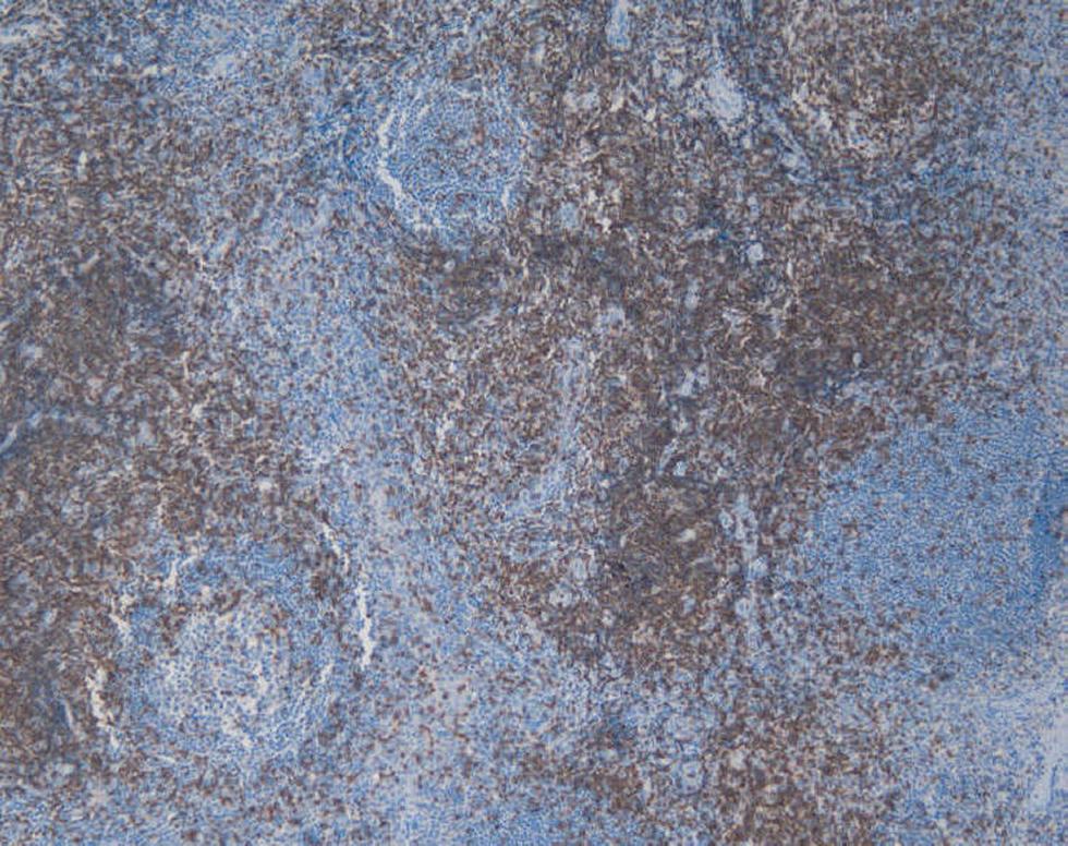
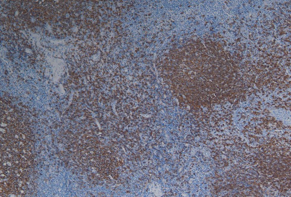
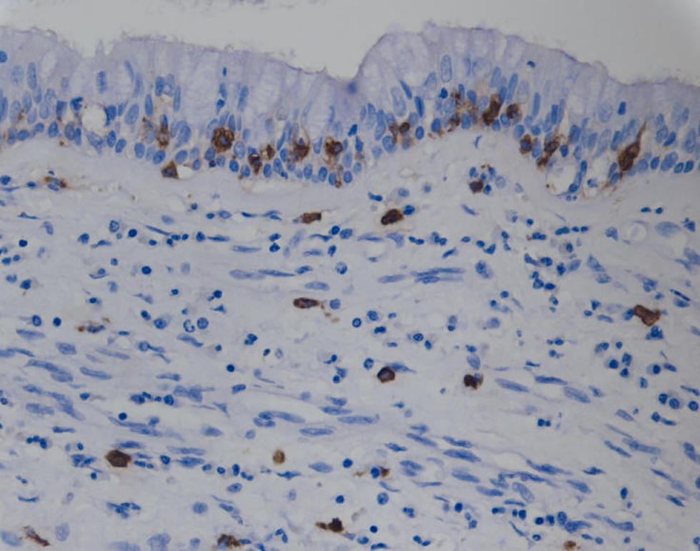
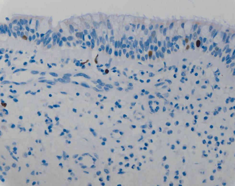
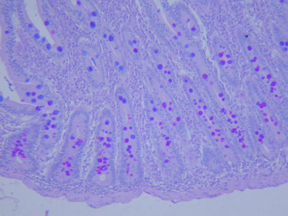
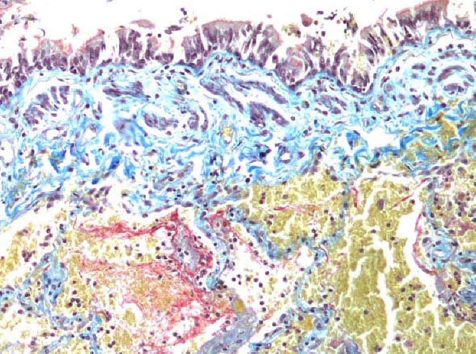
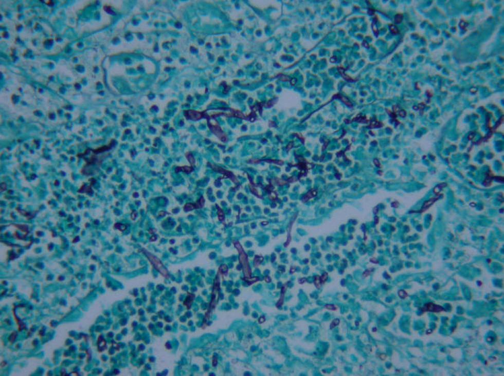
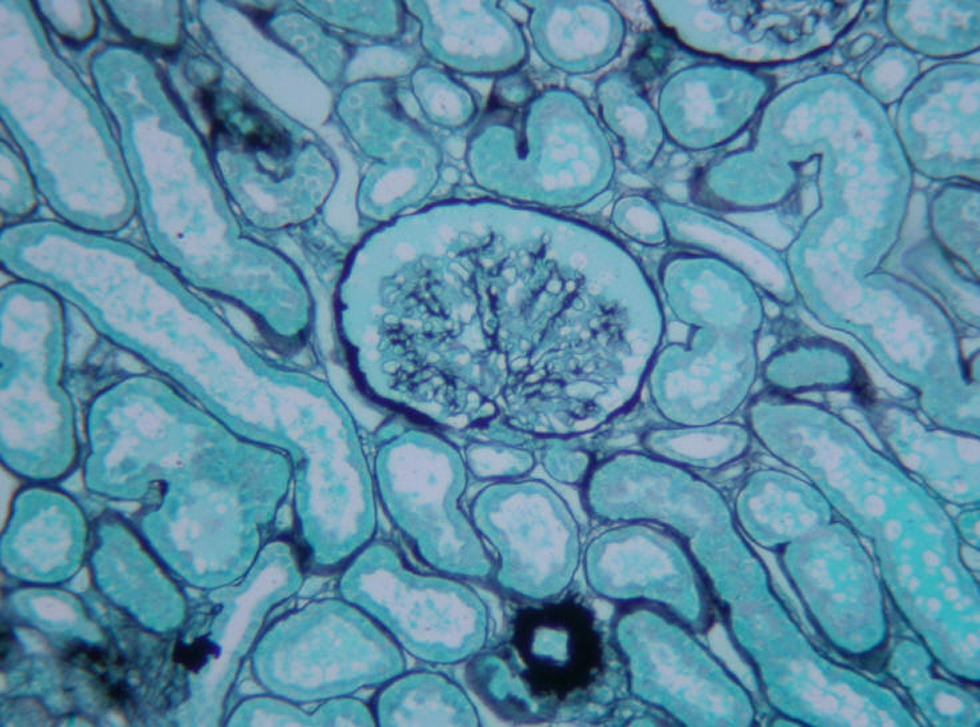
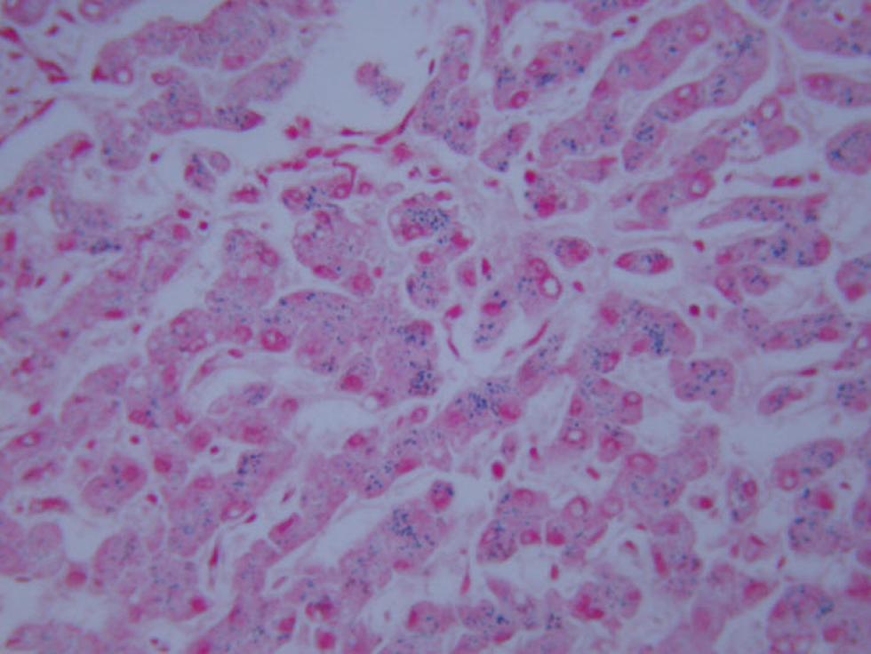
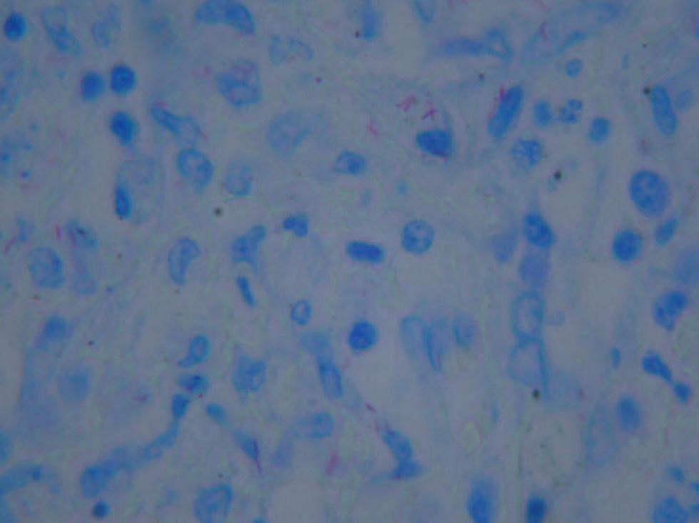
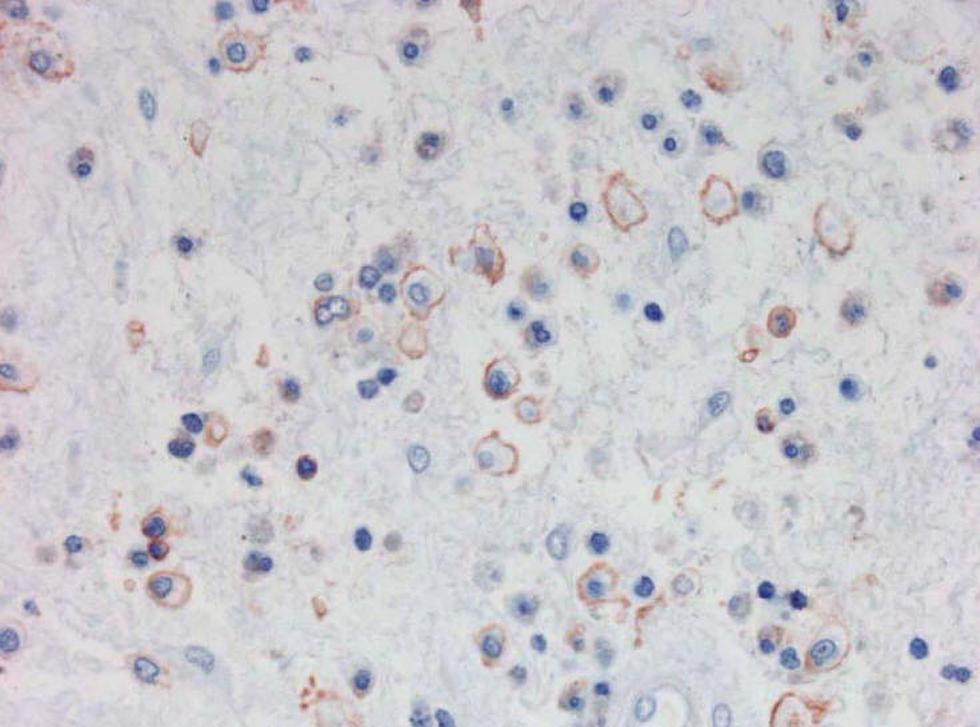
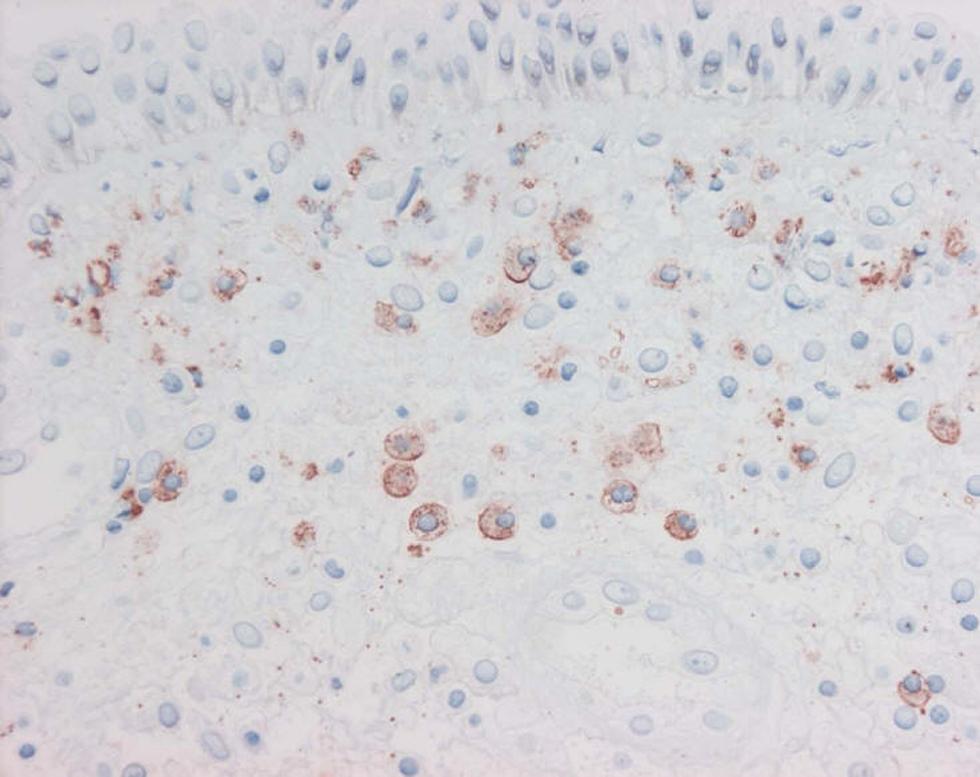
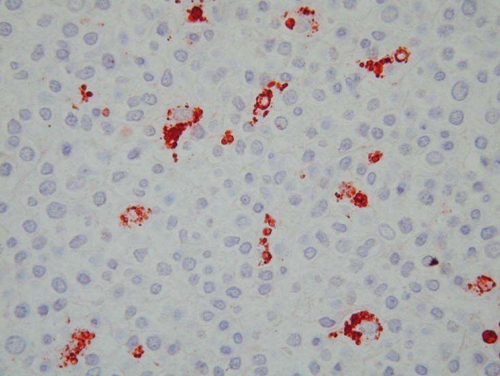
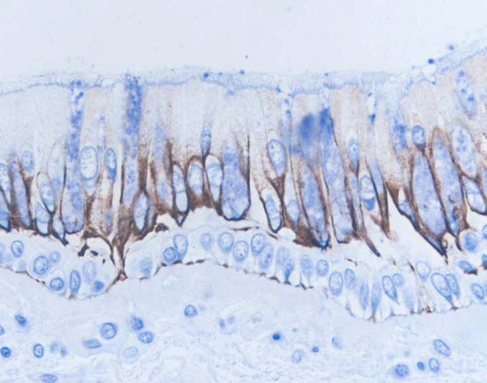
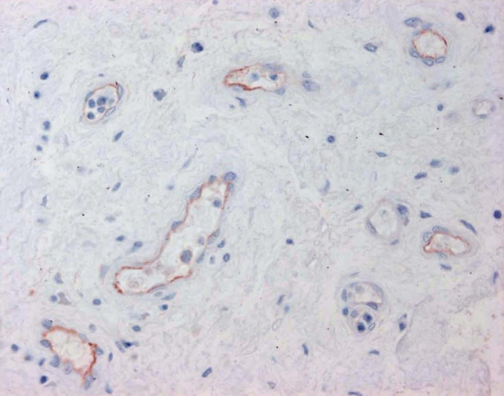
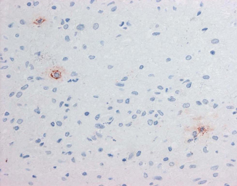
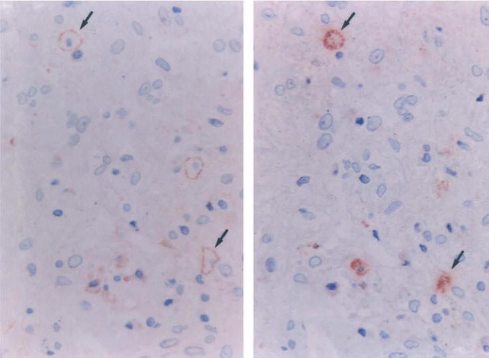
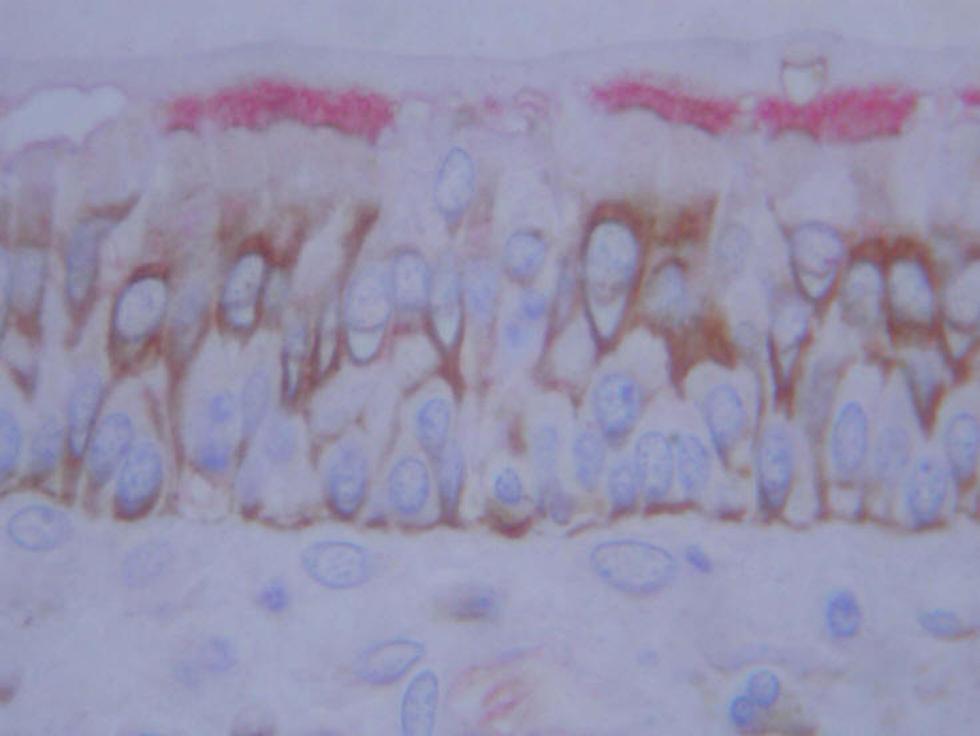
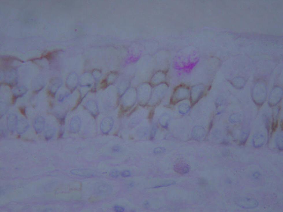
Section of paraffin embedded tonsil immunohistochemically stained (brown) with anti-CD3 to demonstrate T lymphocytes.Section Section of paraffin embedded tonsil immunohistochemically stained (brown) with anti-CD20 to demonstrate B lymphocytes.Section of paraffin embedded nasal polyp immunohistochemically stained (brown) to demonstrate mast cells (AA1+).Section of paraffin embedded nasal polyp immunohistochemically stained (brown) with the nuclear proliferation marker ki67.Paraffin section of jejunum stained by the Alcian Blue / Periodic Acid Schiff technique showing differential staining of mucins. Neutral mucins are pink, acid mucin blue and mixed mucins various shades of purple.Paraffin section of lung tissue stained by the MSB technique. The bronchial epithelial cells are stained purple and overlie the lamina propia within which collagen is stained blue, muscle red and erythrocytes yellow.Paraffin section of kidney stained by the Grocott method to demonstrate the presence of fungi. The hypae of monilia are stained blackParaffin section of kidney stained by the Jones hexamine silver technique for basement membranes. In this section the membranes around the glomerulus, the glomeruli tuft and around the tubules are stained black.Paraffin section of liver stained by the Perls histochemical technique to demonstrate iron (blue).Paraffin section of lung stained by the Ziehl Nielson method to show the presence of tuberculosis bacteria (red).Section of GMA embedded nasal polyp immunohistochemically stained (red) with anti-CD4 to demonstrate T helper cells.Section of GMA embedded nasal polyp immunohistochemically stained (red) to demonstrate eosinophils (EG2+).Section of GMA embedded tonsil immunohistochemically stained (red) to demonstrate macrophages (CD68+).Section of GMA embedded bronchial mucosa immunohistochemically stained (brown) for cytokeratin 18.Section of GMA embedded nasal polyp immunohistochemically stained (red) to demonstrate the vascular adhesion molecule VCAM-1.Section of GMA embedded nasal polyp immunohistochemically stained (red) to demonstrate interleukin-4 (4D9+).Sequential sections of GMA embedded nasal polyp immunohistochemically stained (red) to demonstrate interleukin-4 (3H4+) and mast cells (AA1+), respectively. Several cells are stained with both antibodies.Section of GMA embedded nasal polyp double stained with an antibody to CBE-1 (brown) and an antibody to b tubulin (pink) [Yoshisue H et al].Section of GMA embedded nasal polyp double stained with an antibody to CBE-1 (brown) followed by the periodic acid schiff technique (pink) [Yoshisue H et al].