Improving imaging at an atomic level

Life scientists will meet at the University of Southampton later this month to explore how researchers from different disciplines can work together to improve the accuracy of the imaging of biomolecules. This could help pave the way for the development of advanced pharmaceutical drugs.
More than 60 delegates, largely from the south of England, will examine how a combination of laboratory experiments and computational models can enhance our understanding of large biomolecules and molecular machines.
The symposium has been organised by Jon Essex, Professor of Computational Systems Chemistry and Dr Ivo Tews, Lecturer in Structural Biology.
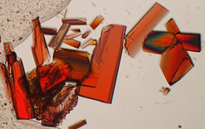
While it is possible to view biomolecules at the atomic level, for example using x-ray crystallography and nuclear magnetic resonance spectroscopy (NMR), there is a revolution about: the latest generation of electron microscopes have become extremely powerful, and interpretation through computer simulation is leaping forward. Using a combination of experimental and computational techniques helps to understand how biomolecules perform the essential functions of life.
“Together with development of new nanocrystal X-ray crystallographic techniques and the advent of free electron lasers (XFEL), we can start to visualise how molecules change over time”, says Ivo.
“If we know more about how molecules fit together, it could eventually lead to more opportunities for colleagues working in pharmaceutical research to develop new drugs,” says Jon.
Both scientists are members of Southampton’s Institute of Life Sciences.

