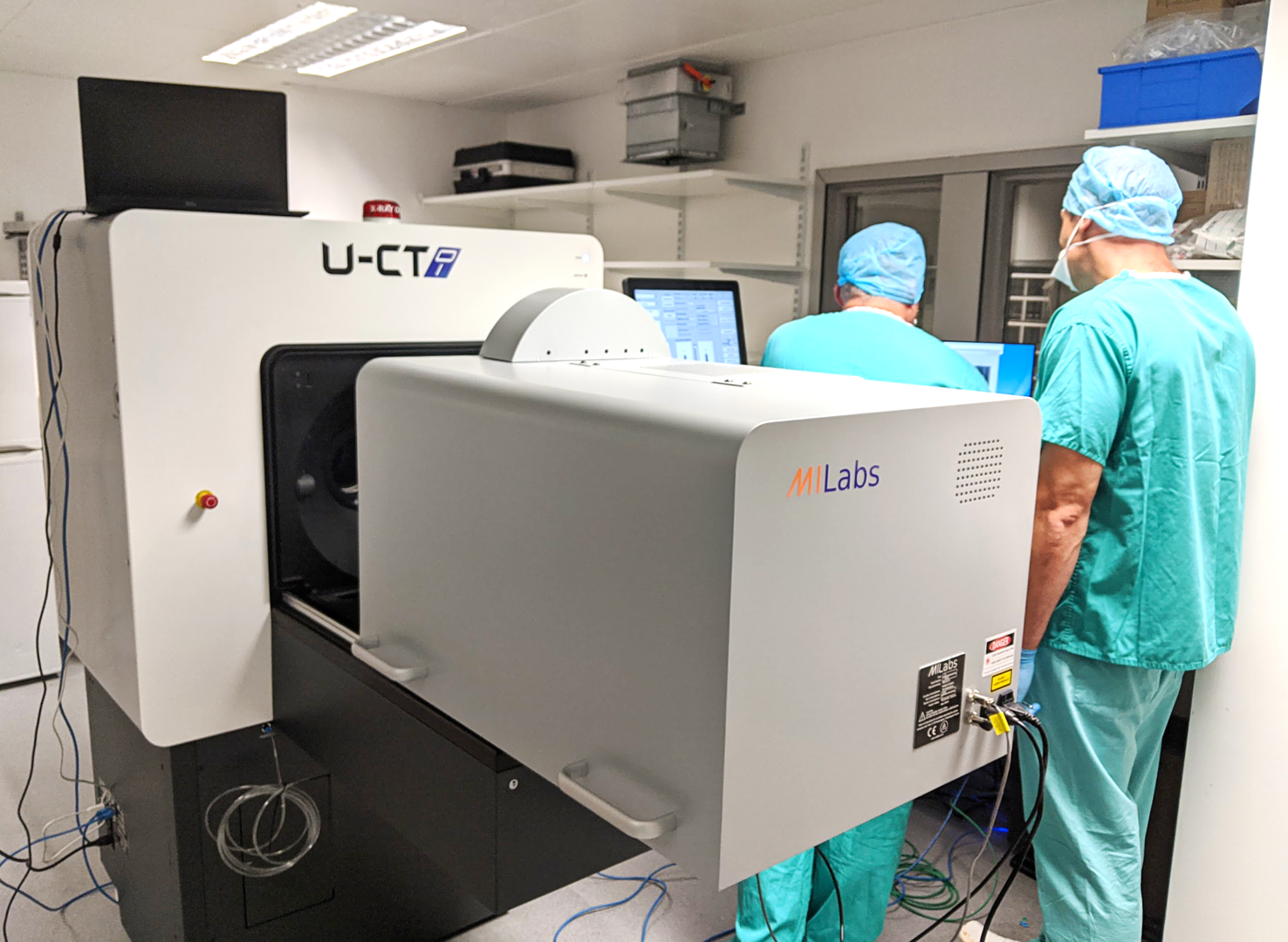
An in vivo small animal dual imaging system to track the spatial and temporal distribution of tissue structures and physiologically relevant labels (bone and soft tissues including lung, liver, adipose tissue, vascular systems and cancerous tissue). The system is hosted at University of Southampton's research facilities at Southampton General Hospital and primarily run by Dr. Katie Dexter and the BIU.
In a nutshell:
‣ Ability to scan living organisms up to the size of a small rabbit (up to five mice can be scanned at once).
‣ Correlative imaging to map fluorescence/luminescence images onto X-ray CT images and white light images.
‣ Data manipulation via MiLabs software and/or third party software.
µCT modality
‣ Spatial resolution down to c. 13 μm
‣ 3D fluorescence, bioluminescence and Cherenkov luminescence tomography
Optical system
‣ Resolution of c. 3 mm
‣ Visual to near-IR range (340 nm to 830 nm; up to six emission filters and up to 11 excitation filters; C-MOS detector)
‣ Dyes are separated using spectral unmixing.