People
μ-VIS Staff and Academic affiliates
A Centre for Computed Tomography at Southampton
serving the Biomedical, Engineering, Environmental and Archeological sciences
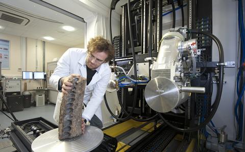
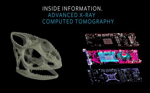
www.muvis.orgThe µ-VIS X-Ray Imaging Centre is University of Southampton's dedicated centre for microfocus Computed Tomography (µCT) and founding partner of the National Research Facility for lab-based X-ray CT (NXCT). The centre combines state-of-the-art equipment and 25 years of experience, plus the expertise of over 40 academic staff from across the university. µ-VIS constitutes a strategic multimillion pound investment (>£5M) in high resolution X-ray tomographic imaging, offering a unique user experience for advanced 3D imaging. We specifically aim to provide a holistic approach to advanced 3D (volume) imaging, supporting all steps from hypothesis formation and experimental design, through to verified, publishable conclusions.

state-of-the-art X-ray tomographic imaging facilities for industry and academia
NXCT is the UK’s National Research Facility for lab-based X-ray Computed Tomography.
Funded by the Engineering and Physical Sciences Research Council (EPSRC), our mission is to provide access and expert support for both academia and industry, embracing both first-time users and more experienced researchers to run cutting-edge 3D and 3D time-lapse imaging experiments.
Launched in November 2020, NXCT is investing in new CT scanning capability to build on existing facilities and expertise – from the Universities of Manchester, Southampton, Warwick, University College London (UCL), and Diamond Light Source – to deliver unparalleled 3D imaging capability for the UK.
Related articles:
NXCT Homepage | Our contribution | Details of the EPSRC Grant
μ-VIS Staff and Academic affiliates
Maps / directions to the facility, postal address for specimens and email address for enquiries
Scan request forms, Charges and Access options for Academic and Industrial users
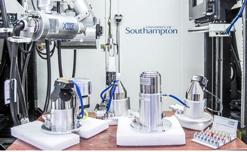
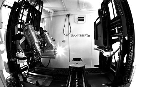
Complementary X-CT systems supporting a range of sample sizes and spatial resolutions as well as in situ experiments

µ-VIS organises training sessions on a regular basis and occasional workshops on more specialised topics

Latest stories of µ-VIS activities
Virtual reality tours of µ-VIS facilities at Highfield campus
µ-VIS contribution to a number of representative projects
Photorealistic 3D volume rendering videos
Information about µ-VIS
|
Announcements
|
|---|
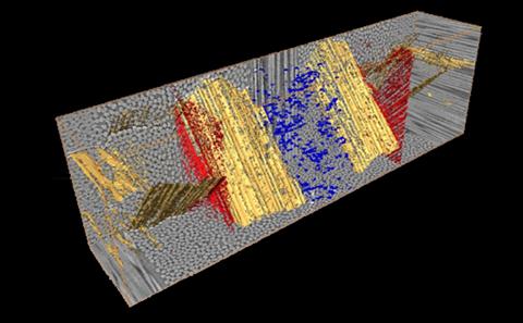
Setting the foundations for X-ray micro-computed tomography workflow for non-destructive 3D X-ray histology (XRH): X-ray imaging by micro-computed tomography (uCT) is a key technique in the rapidly developing field of 3D microscopy. Until recently, 3D imaging of wax-embedded histological tissues was beyond the reach of microCT. However, we have shown that it is possible to non-invasively generate microstructural detail from standard tissue blocks and have provided evidence that this can overturn misconceptions of disease development when just relying on 2D assessment. Building on our previous Wellcome Trust Pathfinder funding, we will develop a research capability, emphasising X-ray hardware performance and flexibility, large-scale computing resource/tools and validation with 2D histology.
www.xrayhistology.org