What is 3D X-Ray Histology (XRH)
A technique tailored to fit seamlessly into current histology workflows in biomedical and pre-clinical research, as well as clinical histopathology
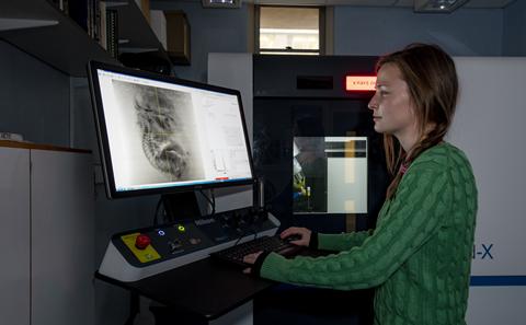
X-ray microfocus Computed Tomography -based workflow for non-destructive 3D X-ray histology (XRH)
μ-VIS X-Ray Imaging Centre | Biomedical Imaging Unit Funded by Wellcome Trust
www.xrayhistology.orgRedirecting...
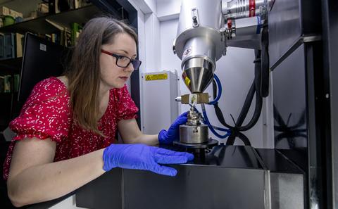
Welcome to the 3D X-ray Histology (XRH) Facility at the University of Southampton, a pioneering platform in the field of histology and biomedical imaging. In collaboration with the University Hospital Southampton (UHS) NHS Foundation Trust and industrial partners, our facility specialises in 3D imaging workflows using X-ray microfocus computed tomography (μCT). Uniquely designed with primary focus on whole-block XRH imaging of tissue specimens, we offer correlative imaging with techniques like classical histology, immunohistochemistry and electron microscopy.
From user support to data analysis, training, and metadata generation, the XRH facility covers every aspect of the imaging process.
Find out more at:
Katsamenis OL, Basford PJ, Robinson SK et al. A high-throughput 3D X-ray histology facility for biomedical research and preclinical applications, Wellcome Open Res 2023, 8:366, https://doi.org/10.12688/wellcomeopenres.19666.2
Foundations for routine 3D X-ray histology (XRH) project
On 2018 our team was awarded an £1.14M follow-up* Wellcome Trust, Biomedical Resource and Technology Development grant to further develop a flexible technique for non-destructive 3D (volume) imaging and analysis of standard tissue samples via X-ray micro-computed tomography (µCT).
The the technique, which we identify as 3D X-ray Histology (XRH) is integrating precise registration and correlation with conventional histopathology and will support high-throughput imaging of routinely prepared formalin-fixed and paraffin embedded tissue specimens and standarised and automated workflows, through new X-ray equipment. We envisage this to be directly translated into next-generation clinical image-based diagnostics and patient stratification using artificial intelligence and deep learning
* (2016-2017) Wellcome Trust, Pathfinder Award, “Development of micro-computed tomography (μCT) for enhanced diagnosis and prognosis in interstitial lung diseases (ILD)”
A technique tailored to fit seamlessly into current histology workflows in biomedical and pre-clinical research, as well as clinical histopathology
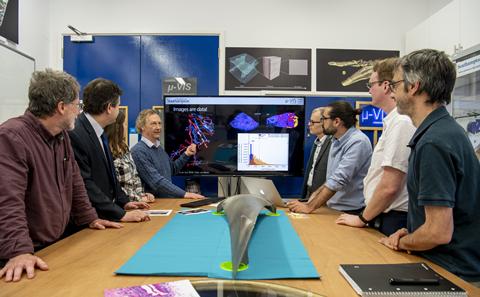
Meet the team, and the laboratories involved
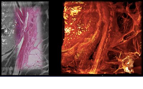
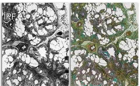
Representative application of 3D X-Ray Histology
Read about the journey to this point and our vision for the future of XRH

List of conference and journal publications
Find out how you can work with us and see what XRH can offer to your research
A centre for Computed Tomography at Southampton, serving the Biomedical, Engineering, Environmental and Archeological sciences
Research and diagnostics high resolution light and electron microscopy facility, with a special emphasis on 3D imaging.