https://www.toscainternational.org/tosca-UK-2019
Orestis L. Katsamenis1*, Christina Karavasili2, Dimitrios G. Fatouros2
1 μ-VIS X-Ray Imaging Centre, Faculty of Engineering and the Environment, University of Southampton, SO17 1BJ Southampton, UK;
2 Laboratory of Pharmaceutical Technology, School of Pharmacy, Aristotle University of Thessaloniki, GR-54124, Thessaloniki, Hellas;
Combination chemotherapy is currently arising as means of overcoming multidrug resistance and enhancing therapeutic response, while reducing administered drug dose and side effects. The term refers to the delivery method that utilises the synergistic (or additive) effect of multiple anticancer agents in a co-delivery formulation. Doxorubicin (DOX) is an anticancer agent used to treat a wide range of cancers and despite its high toxicity it remains in use because of its efficacy in the treatment of several types of tumours [1]. Curcumin (CUR) is a polyphenol originating from the rhizomes of turmeric and has been broadly evaluated for its anticancer potential [2]. The utilisation of nutritional constituents in therapeutics, often referred to as Nutraceuticals, encompass multitargeting efficacy and lower toxicity compared to the conventional synthetic moieties.
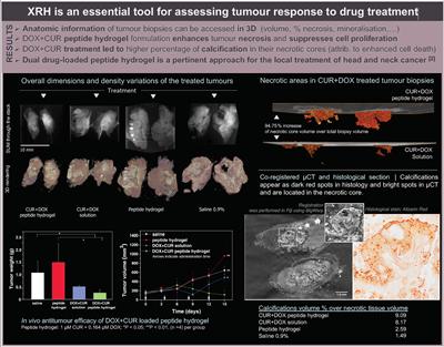
In this study we have synthesised and assessed the therapeutic potency of the ac-(RADA)4-CONH2 self-assembling peptide hydrogel for the co-delivery of DOX and CUR against head and neck cancer cells in HSC-3 cell-xenografted severe combined immunodeficient mice. 3D X-ray histology (XRH)* was employed to provide high-resolution 3D (volumetric) anatomic information of tumour response after treatment by analysing the phenotype of the excised tumours (figure). XRH imaging of the excised tumours allowed us to quantitively analyse the volumetric characteristics of anatomical features, such as tumour volume, its necrotic core and calcifications.
Investigation of the ratio between the total tumour volume and the necrotic tissue volume within them highlighted the efficacy of the DOX+CUR peptide hydrogel to enhance tumour necrosis and suppress cell proliferation. Our study demonstrates that 3D X-ray histology is a powerful tool for providing high-resolution anatomic information of tumour biopsies after intratumoral administration of chemotherapeutic formulations.
* 3D X-ray histology (XRH) is a µCT-based workflow for imaging unstained paraffinized tissue specimens developed in the University of Southampton, tailored to fit seamlessly into current histology workflows [3].
[1] P. K. Singal and N. Iliskovic, "Doxorubicin-induced cardiomyopathy", New England Journal of Medicine, vol. 339, no. 13, pp. 900-905, 1998
[2] R. Wilken, M. S. Veena, M. B. Wang, and E. S. Srivatsan, "Curcumin: A review of anti-cancer properties and therapeutic activity in head and neck squamous cell carcinoma", Molecular cancer, vol. 10, no. 1, p. 12, 2011
[3] O. L. Katsamenis et al., "X-ray micro-computed tomography for non-destructive 3D X-ray histology", The American journal of pathology, 2019