Aileen O’Brien1, David I. Whiteside2 and John E. A. Marshall1
1School of Ocean and Earth Science, University of Southampton, National Oceanography Centre Southampton, European Way, Southampton, SO14 3ZH, UK
2School of Earth Sciences, University of Bristol, Bristol, BS8 1RJ, UK
Background
Computed tomography is increasingly being used as a non-destructive, investigative tool in palaeontology, yielding valuable information about unseen portions of fossils, partially or wholly embedded in rock matrix.
Role of µ-CT in our study
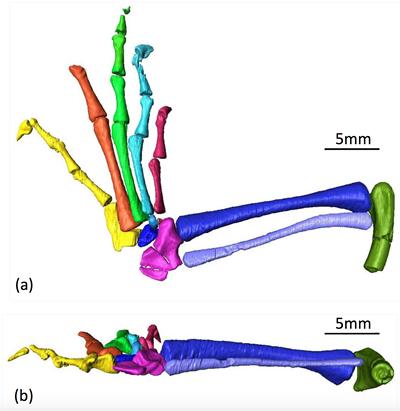
µ-CT images of a partially complete, left hind limb of Clevosaurus hudsoni (NMHUK PV R36846) were obtained at µ-VIS and the data processed using Avizo 3D visualisation software, in the µ-VIS visualisation suite. The surface models created from the µ-CT scans provide a more complete picture of the morphology of the hind limb, including non-visible parts of the bones concealed within the matrix. Features of the bones such as compression in a particular plane, grooves, processes, facets and projections are often disguised by matrix, or are hidden by adjacent bones. Surface modelling of µ-CT data has highlighted these characters and yielded insights into the ontogeny and age of the animal from which the specimen came.
Acknowledgements
The authors would like to thank Sandra Chapman (NHMUK) for arranging the loan of Clevosaurus hudsoni.