Immunohistochemical staining
Immunohistochemical staining
We have over 20 years experience in the use of immunohistochemical techniques. The HRU has a range of validated protocols suitable for frozen, paraffin and GMA resin sections.
We stock a broad range of primary antibodies including those to identify cells, adhesion molecules, epithelial components, assess cell proliferation status and cytokines optimised for the study of inflammatory processes in a variety of tissues.
The HRU also stocks a range secondary antibodies, detection kits and chromagens.
We have optimised double staining using either two primary antibodies or an antibody in combination with a tinctorial stain.
Immunohistochemical staining
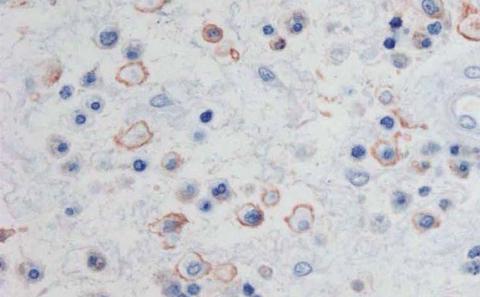
GMA section of nasal polyp stained by immunohistochemistry for CD4+ lymphocytes
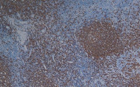
Paraffin section of tonsil stained by immunohistochemistry for CD20+ lymphocytes
![GMA section of nasal polyp double stained for beta tubulin (pink) and CBE-1 (brown) [Yoshisue H et al]](https://cdn.southampton.ac.uk/assets/imported/transforms/content-block/BoxGridWithImages_ImageBlock/69718A4D0FCC456EA37BF176FDCDD400/img19_cropped.jpg_SIA_JPG_fit_to_width_MEDIUM.jpg)
GMA section of nasal polyp double stained for beta tubulin (pink) and CBE-1 (brown) [Yoshisue H et al]