Andreu Laborda, Songsong Lu and Philippa Reed
Materials Group, Mechanical Engineering Dept, Faculty of Engineering and Physical Sciences, University of Southampton
Background
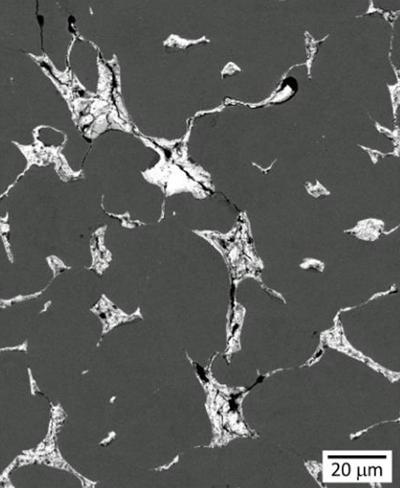
In multilayer plain journal bearings, leaded bronze forms the interlayer that is bonded to the steel-backing and then coated by thin overlay coatings. In general, it needs to support the cyclic loads transferred from coatings, which provide the desired material properties to ensure the requirements of hydrodynamic lubrication. In addition, it also needs to serve as a solid lubricant by smearing out lead to bear loads transferred from the oil film in cases where the coatings are removed from the bearing surface under certain boundary lubrication conditions. Moreover, replacing the lead in this kind of material system to reduce pollution is a key focus. Thus understanding the inherent fatigue performance of dissimilar materials systems comprising complex 3D interlinked phases with very different mechanical properties (such as the leaded bronze) is both challenging but also has clear industrial research relevance.
Role of Computed Tomography
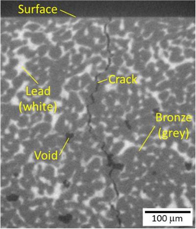
The micro-CT capabilities at µ-VIS can produce high resolution 3-dimensional data of the internal structure of the material, which can be applied to understand the cracking processes in the leaded bronze material system. It allows capture of the 3D characteristics of the lead phases, bronze matrix and the crack growth plane. The micro-CT data have also been employed to build a RVE model to develop our understanding of the failure in complex inhomogeneous two phase systems with dissimilar material properties.
Acknowledgements
The authors would like to thank Dr. Daniel Bull at University of Southampton for his assistance with image processing, Grazina Burmistroviene and Guy Lowmann for assistance on sample preparation and manufacturing. This work was supported by the Engineering and Physical Sciences Research Council (EPSRC, EP/M50662X/1) and Daido Metal Co. Ltd.
Figure 1 - SEM observation of crack propagation in the cross-section of leaded bronze
Figure 2 - µ-CT slice of a crack propagating through the interlayer