

By combining patient-derived tumour models, advanced molecular analysis and AI-supported decision-making, Tim's team is helping clinicians understand how a cancer is likely to respond before treatment begins. His work brings together surgeons, scientists and computational experts to create a future where care is more precise, more personal and gives more patients the best possible chance of a better outcome.
Precision oncology has become increasingly sophisticated at describing cancer. The harder challenge is turning biological complexity into a better decision for the patient in front of the clinical team.
That challenge sits at the centre of Tim’s research. Together with colleagues in the Innovation for Translation Research Group (ITRG), he is working towards a model in which clinical insight, molecular analysis, patient-derived tumour models and artificial intelligence combine to support more individualised treatment decisions.
Tim and the ITRG's ambition is not simply to give a better standard of care, but better evidence for determining the right care for an individual patient.
“AI is central to our approach, but not as a replacement for clinical expertise,” says Tim.
Concentrating expertise where it is needed most
Cancer multidisciplinary teams (MDTs) must interpret increasingly complex clinical, imaging, histological and molecular information while managing a growing caseload.
“We explore whether machine learning can make that information more tractable and help clinicians concentrate their expertise on the cases that most require it,” he adds.
The team’s research in oesophageal cancer demonstrates the potential of this approach.
In collaboration with Professor of Artificial Intelligence, Gopal Ramchurn, the team used explainable artificial intelligence (XAI) to quantify how specific clinical variables contribute to MDT treatment decisions. Importantly, clinicians not only get an output, but sufficient transparency to interrogate it and exercise independent judgement.
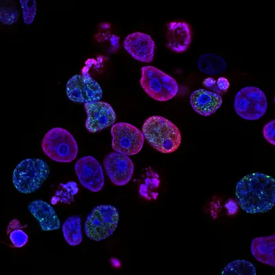
This computational strand is complemented by sophisticated experimental modelling, which is illustrated by a recent collaboration with the Sanger Institute.
"We were involved in a major programme establishing 256 clinically annotated patient-derived tumour organoids across colorectal, oesophageal, ovarian, pancreatic and gastric cancers.”
Tim Underwood, Professor of Gastrointestinal Surgery
“Whole-genome and transcriptome sequencing was combined with genome-wide CRISPR-Cas9 screening of 162 organoids, creating a functional map of cancer gene dependencies.”
The distinction is important.
Genomic profiling can identify what is altered in a tumour; functional perturbation can reveal what that tumour depends on.
Using MinLibCas9 and Yusa v1.1 libraries, with analytical methods including CRISPRcleanR and BAGEL2, the team was able to identify context-specific vulnerabilities and integrated them with genomic, transcriptomic and clinical characteristics.
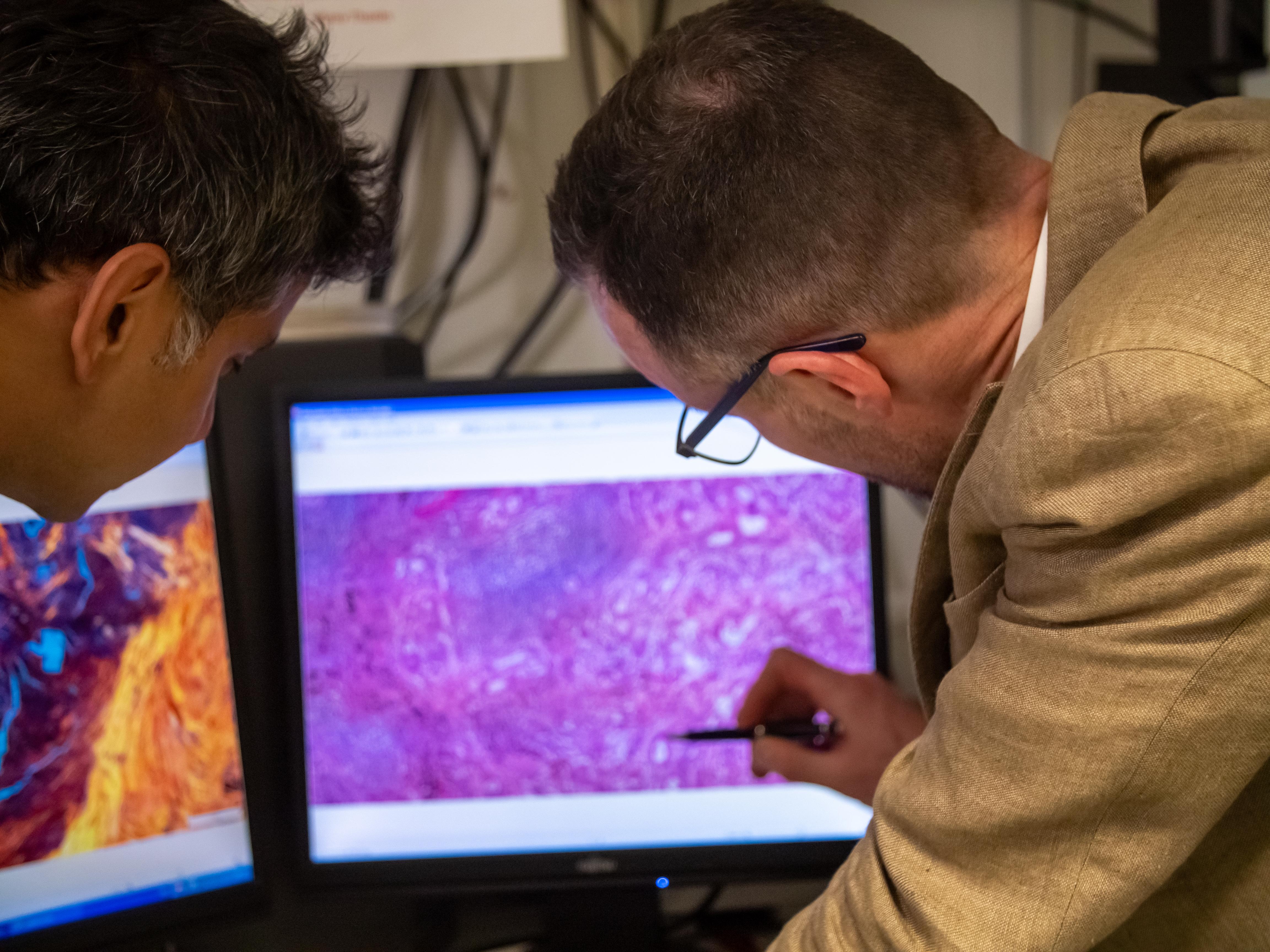
Combining disciplines to tackle the problem
“The approach was backed with using real patient samples collected before treatment. Following relapse also exposed changes in gene dependencies and drug sensitivity associated with tumour evolution under therapeutic pressure,” says Tim.
The significance extends beyond individual targets. This is an approach that connects computational prediction with experimental biology rather than treating either discipline as sufficient on its own.
“Patient-derived organoids, multi-omics, CRISPR perturbation and explainable AI provide different layers of evidence that can potentially inform the same clinical problem,” he adds.
For gastrointestinal cancer research, this reframes the central question. Not simply: what type of cancer is this? But: what does this particular cancer depend on, how is it changing, and which evidence can best support the next clinical decision?
That is the compelling direction in Tim’s work: using AI not to replace human judgement, but to give it a richer and more individual evidence base.
If we can combine advanced modelling, AI, molecular analysis and clinical expertise to understand each person's cancer in greater detail, we can move beyond treating averages and start treating individuals. That's the future I want to help build. Not a better standard of care for everyone, but the right care for each person.