
About
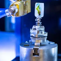
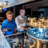
The Ultrafast X-ray group works on the generation and uses of extreme ultraviolet (XUV) and soft X-ray radiation using high energy femtosecond laser pulses. The X-rays are produced using high harmonic generation (HHG), a process which uses electron ejection and recombination to produce very short (attosecond) pulses of light.
Soft X-ray radiation has many important uses. Its short wavelength makes it ideal for high-resolution microscopy. Many chemicals have absorption edges in the soft X-ray region, making it an important area for spectroscopy.
Finally, the time resolution available from the generated pulses gives us the overall possibility of femtosecond time resolution and nanometre spatial resolution within the same instrument, which opens whole areas of exciting new science.
The research effort in this area is multi-disciplinary. Within the ORC we collaborate with Dr Peter Horak, an expert in nonlinear optics. The group has strong collaborations with the School of Chemistry (Prof Jeremy Frey, Dr. Russell Minns), where X-ray scattering using more traditional sources is a long-standing and prestigious research area (the National Centre for X-ray crystallography is based in Southampton Chemistry). More recently, we are building collaborations with the Institute for Life Sciences in biological imaging (John Chad, Katrin Deinhart), and the Rosalind Franklin Institute.
Key to the process of soft X-ray microscopy with HHG sources is the use of phase retrieval techniques. These are computer algorithms that allow us to generate an image without the use of an objective lens. The scattered light from the illuminated object is collected, and the phase information necessary to recreate an image of the object, lost during the collection process, is recreated using an iterative algorithm. These algorithms can now be combined with machine learning to create new imaging techniques on the nanoscale.
People, projects, publications and PhDs
People
-
Bill Brocklesby
Novel imaging and microscopy techniques in the visible and XUV spectral regions;
Email: wsb@orc.soton.ac.ukTel: +44 23 8059 2042
Related research institutes, centres and groups
-
Institute for Life Sciences
We bring together interdisciplinary researchers with expertise across the themes of health and medicine, living systems, disruptive life technologies, and insights through data -

Statistical Sciences Research Institute (S3RI)
We use statistics and demography methodologies to improve the way data is used. -

Computational Systems Chemistry
Our research covers a broad range of domains from nanoscale materials properties, atmospheric chemistry, through to biological systems. -
Southampton Imaging
Imaging has become an essential part of scientific research, from biomedical sciences to engineering to optoelectronics.
Connect with us
Enquiries
Optoelectronics Research Centre
University of Southampton
Southampton
SO17 1BJ