

For more than a century, we have known that centrosome abnormalities are a hallmark of cancer. Tumour cells often carry too many of them, which disrupts chromosome segregation and causes aneuploidy and genetic instability. But a basic question has remained open: are all centrosome abnormalities the same, or do different kinds drive different aspects of the disease?
The obstacle was scale. For decades, studying centrosomes meant counting them by hand. Even at a microscope this was painstaking: each one is a tiny dot inside a cell, and a single tumour sample can hold thousands. Semi-automated tools have helped in recent years, but they cannot capture a tumour’s wider spatial context, and human variability and error remain a problem. As a result, studies have stayed small.
Until now.
Dr Salah Elias, Associate Professor in the School of Biological Sciences, and colleagues have developed an AI platform called CenSegNet, capable of analysing whole tumour samples, cell by cell, at speed with precision.
It has revealed that two defects in centrosomes, long treated as one, behave very differently, which could change how breast cancer is treated in the future.
“Studying centrosomes at scale has been challenging because of the limited availability of high-throughput quantitative tools. By combining these tools with imaging, we can gain a clearer picture of how centrosome abnormalities relate to cancer.”
Dr Salah Elias
Innovative collaborations
Salah’s lab studies how cells in the breast decide which way to divide, which shapes how a tissue grows and what happens when that goes wrong. Centrosomes steer that decision, so the team needed to measure centrosomes accurately across many samples. They expected that to be the easy part.
“We didn’t see it as a technical problem,” Salah says. “We thought, as cell biologists, we just need to count them and that’s it, because we wanted to address a bigger biological question.”
It turned out to be far harder than that, and it set the project on a new course.
They turned to computer scientists and AI experts at Southampton to adapt and combine existing computer vision methods for detecting and outlining centrosomes in microscopy images.
The resulting platform, CenSegNet, combines two algorithms: one locates centrosomes, while the other traces their outlines to measure their size and shape. “It’s like cooking,” Salah explains. “You have different ingredients, each of them good, but they do not fulfil everything in the dish, and the AI still needs human teaching. It’s very powerful and allows you to do this at scale, but you need to train it,” he says. “You need scientists who know the biology and are willing to annotate thousands of centrosomes.”
Jiaoqi Cheng, a PhD student on the project, did exactly that, by carefully marking centrosomes by hand in tissue images to provide reference annotations for training the system. “You need accurate, very good human input before you train these algorithms to be highly accurate,” adds Salah.
The team also trained it on images from several kinds of microscopy, including pathology imaging, high-resolution imaging and super-resolution imaging. That way, other labs can use the same pipeline with whatever equipment they have. It also had to learn to read each centrosome in context, because the tissue around it is complex and varies from one region to the next.
Two defects, not one
Once formed, CenSegNet was used to analyse more than 330,000 centrosomes across 911 tumour specimens from 127 patients, in collaboration with University Hospital Southampton.
It uncovered two distinct forms of centrosome abnormality that had previously been considered part of the same process. One form involved cells acquiring too many centrosomes, while the other involved abnormally enlarged centrosomes. Surprisingly, the researchers found that these defects behave independently and can occupy different regions of a tumour.
The study also revealed important links between centrosome abnormalities and clinical features of disease. Tumours with high levels of enlarged centrosomes were associated with more aggressive characteristics, including higher tumour grade, lymph node involvement and certain genetic alterations. Importantly, patients whose tumours contained fewer enlarged centrosomes in the tumour core tended to have better overall survival.
“Previous studies have not always distinguished between these different types of centrosome abnormality. With CenSegNet, we can distinguish numerical from structural centrosome abnormalities and investigate how each is associated with different features of breast cancer.”
Dr Salah Elias
What it could mean for patients
CenSegNet is currently a research tool rather than a clinical test, but the findings suggest several possible future applications.
First, spatial maps of centrosome abnormalities could help researchers assess whether these patterns provide additional information about tumour aggressiveness and patient outcomes beyond established clinical measures. Larger, independent studies would be needed before they could guide patient care.
Second, the work could inform research into treatments that target centrosome-related vulnerabilities. Distinguishing between different defects may help researchers investigate which tumours could respond to such approaches, although the study did not establish a way to match patients to treatments.
Third, the work provides a framework for studying how centrosome abnormalities vary between regions of the same tumour. This could help researchers investigate how these differences relate to tumour progression, spread and treatment response.
CenSegNet is freely available as open-source software, and Salah hopes cancer researchers and pathologists around the world will use it in their research. Beyond breast cancer, the team has demonstrated its use in other tissues, including kidney, colon and appendix samples.
More broadly, the work shows how AI can help researchers study individual cellular structures within their tissue context, opening up new ways to investigate their links to disease.
Why Southampton?
Salah says the project brought together a combination of expertise and resources that is not available everywhere: cell biology, patient tissue, computer science and AI. Southampton’s interdisciplinary culture and the community’s willingness to work together helped him find the expertise the project needed.
Cancer scientists and clinicians provided access to patient samples, while computer scientists brought expertise in image analysis. A small pilot grant from the Institute for Life Sciences provided early support for the collaboration.
“It was a small grant, but it helped us get the collaboration started,” Salah says.
“We encounter this culture of cross-disciplinary collaboration every day, so it feels natural. Bringing these different areas of expertise together was essential to this project.”
Dr Salah Elias
Perhaps most notably, the team had been unable to find an existing tool that could do what they needed.
“We were surprised that nobody has done it before,” he says, given how central the question is to cancer biology. “I kept asking my students to check the literature, but we were unable to find anything.”
What comes next?
CenSegNet now opens up questions the team could not previously explore at this scale.
Salah’s group is investigating why most breast cells carrying an inherited harmful BRCA1 mutation do not become cancerous. Although the mutation is present throughout the body, cancer arises from only a small subset of cells. The team is asking whether normal tissue organisation and developmental processes, such as how cells divide and orient themselves, help act as safeguards.
Using miniature tissue models, the team will investigate how these processes maintain normal tissue organisation and what happens when they are disrupted.
In a related study, they are asking why levels of a protein called Annexin A1, which has been linked to breast cancer biology and outcomes, vary so widely within breast tumours. They are combining this analysis with centrosome mapping to build a more detailed picture of each tumour.
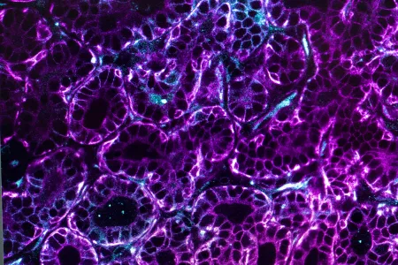
The idea is to adopt a preventive and early detection approach by understanding the initiating events. The longer-term aim is to identify opportunities to detect cancer earlier or prevent it from developing.