About the Biomedical confocal and lightsheet microscopy facility
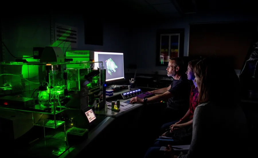
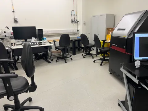
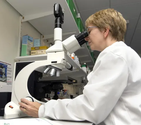
The confocal and lightsheet microscopy facility provides expertise in producing high resolution images.
Confocal microscopy
This uses a pinhole in the light path to discard signal from out of focus regions of a fluorescently labelled (or reflective) sample, thereby generating a sharply focussed, optical section from the sample, without having to physically section it. It is a high magnification, high resolution imaging method which uses a scanning laser beam to excite the fluorescence. Refocussing the microscope and reimaging allows 3D datasets to be collected. Our Leica confocal systems have true spectral detection allowing the simultaneous recording of 4, non-overlapping colour ranges with infinite flexibility and nanometre precision in terms of wavelength detection range.
View our confocal microscopy gallery.
Lightsheet microscopy
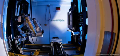
Otherwise known as selective plane illumination microscopy (SPIM), our lightsheet system is a lower magnification, lower resolution method for producing optical sections and 3D images from larger samples (up to 10 x 10 x 5mm). There are many different types of lightsheet systems, using different configurations of light sheet numbers and orientation, providing different degrees of resolution.
It uses a wide, thin sheet of laser light entering the sample from the side (or from multiple sides) to excite fluorescence at a single focal plane which is imaged using a high resolution, ultra sensitive scientific camera. Moving the sample through the laser beam allows signal from different levels within the sample to be captured and 3D data sets generated. Samples for light sheet microscopy often need considerable processing to render them optically clear.
