Biomedical live cell imaging and time-lapse microscopy facility

About the Biomedical live cell imaging and time-lapse microscopy facility

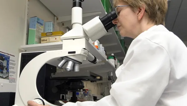
A fully automated, inverted microscope system with brightfield, phase contrast and fluorescence imaging capabilities and long working distance objectives.

The microscope system is housed within an incubator chamber to allow long term observation and recording of cells in conventional microplates, flasks and petri dishes.

Both of our confocal systems are also equipped for live cell imaging and time lapse microscopy, but culture preparation will require different considerations.

Technical specification

Olympus IX81 live cell imaging system

  • fully automated, inverted Olympus IX81 microscope stand
  • Olympus ExCellence software control with graphical programming interface
  • objective range x4 to x60
  • bright field and phase contrast imaging
  • environmental chamber and CO2 controller and premixed 5% CO2 in air feeds
  • standard fluorescence microscopy with FITC/TRITC/DAPI compatible filter cubes
  • electronic stage with relocation function
  • combined phase-fluorescence time lapse facility
  • side and bottom camera ports
  • 1376x1032 pixel high sensitivity Olympus XM10 monochrome camera for time lapse and fluorescence photomicroscopy on bottom port
  • Photron high speed video camera (500 fps) to side port under software control to record bursts of high speed video at specified time intervals

Contact us

Contact us

We are based in the Laboratory and Pathology block at Southampton General Hospital.
We’re open Monday to Friday 09:00 to 17:00 UK time.