About the Biomedical MicroCT facility
Microfocus Computed Tomography (μCT) is a non-destructive imaging technique. It uses X-rays to generate high resolution 3D (volume) datasets from samples. This technique offers spatial resolution down to 6 -8 μm. Able to examine conventional hard, high contrast structures, microCT can also be adapted for imaging softer tissues.
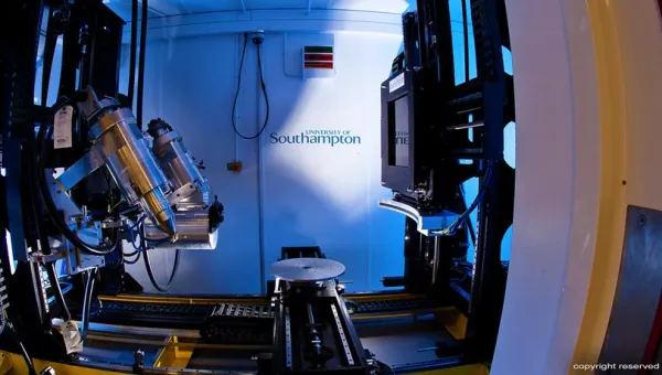
A μCT system consists of a microfocus X-ray source and a high-resolution X-ray detector, enabling imaging at resolutions down to a few micrometers. Depending on the system design, the source and detector can either be mounted on a gantry that rotates around the object or remain stationary while the object is mounted on a rotating stage. The former configuration is typically used in in-vivo systems to accommodate live specimens, while the latter is preferred for ex-vivo imaging, as it offers greater geometric flexibility.