Biomedical scanning electron microscopy facility

About the Biomedical scanning electron microscopy facility

The scanning electron microscopy facility enables the creation of ultra high-resolution images of surface topography with a large depth of focus.

Scanning electron microscopy uses a beam of electrons to scan across the surface of a sample and image the surface topography at high-resolution and with a large depth of focus.

Conventionally, the electron beam interacts with the surface of the specimen, coated with an ultrathin layer of metal, to release secondary electrons which are detected to create the image although backscattered (reflected) electrons from the scanning beam can also be detected.

The electron beam also generates X-rays from the sample which can be detected and characterised to identify and map the elemental composition of the sample. Most imaging is done using specially dried and coated samples under high vacuum, but low vacuum and environmental modes can be used on some uncoated samples.

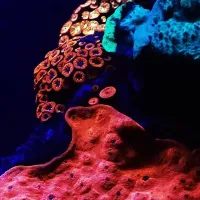
View our scanning electron microscopy gallery.

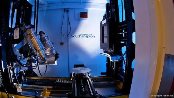
Our ultra high resolution FEI Quanta 250 SEM is equipped with a Gatan 3View II serial block face imaging system for 3D imaging of samples.

Find out more about serial block face imaging.

Technical specification

TESCAN Clara 2 FEGSEM with Katana SBFSEM

  • Ultra High Resolution Scanning Electron Microscope (UHR-SEM) with a Field Emission Gun electron source
  • Secondary electron, Gaseous secondary electron, and backscattered electron detectors
  • Low vacuum mode
  • Montage imaging wit tile stitching
  • Imaging from overview to the nanoscale
  • Integrated Katana Serial Block-Face SEM
  • Volume EM reconstruction software
  • Array tomography capabilities
  • Oxford Instruments Ultim Extreme EDX detector for elemental mapping and analysis

FEI Quanta 250 scanning electron microscope with Gatan 3view2XP serial block face imaging

  • ultra high resolution SEM
  • 30Kv/100,000x magnification
  • Field emission gun
  • High vacuum, low vacuum and environmental modes
  • Secondary electron and concentric ring backscatter detectors
  • Large field and gaseous secondary electron detectors
  • Digital imaging with auto montage facility
  • Beam deceleration to enhance surface imaging
  • Gatan 3View2XP automatic serial block face imaging down to 25nm slices and near TEM resolution (<2nm)
     

Scanning electron microscopy sample processing

  • processing laboratories with fume cupboard, balance, sample rotators, etc.
  • Balzers CPD 030 critical point drier
  • Quorum Q150T ES carbon and sputter coater

Contact us

Contact us

We are based in the Laboratory and Pathology block at Southampton General Hospital.
We’re open Monday to Friday 09:00 to 17:00 UK time.