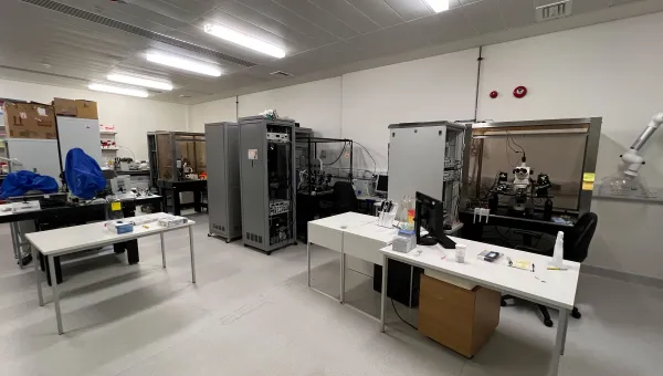
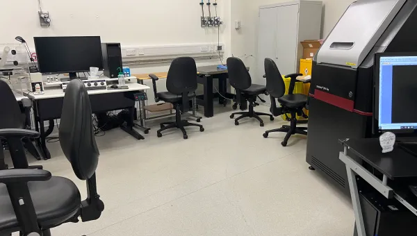
Technical specification
Our widefield and zoom fluorescence microscopes permit the specific labelling of up to four targets simultaneously. We also offer conventional brightfield microscopy to very high magnifications, or on our zoom microscope.
Olympus IX83 Widefield epifluorescence microscope
- epifluorescence - 250 nm resolution
- phase contrast - 250 nm resolution
Objectives
Magnification, numerical aperture and immersion
- 4x, 0.13, air
- 10x, 0.5, air
- 20x, 0.7, air
- 40x, 0.9, air
- 63x, 1.42, oil
- 100x, 1.49, oil
Specimen format
- standard glass slide
- 35 mm coverslip bottom dish
- coverslip bottom chamber-slide
- optical bottom multi-well plate
Detector
Hamamatsu Orca Flash4.0 sCMOS. 2048 x 2048 pixels.
Filtersets
Expansion, electron microscope and colour
- 350/50 nm, 460/50 nm, Blue
- 470/40 nm, 525/50 nm, Green
- 545/30 nm, 620/60 nm, Orange
- 560/40 nm, 630/75 nm, Red
- 620/60 nm, 700/75 nm, Far Red
Other features
- transmissed light - yes
- DIC - no
- environmental temperature/gas control - no
- hardware autofocus - no
- simultaneous acquisition - no
- sequential fluorescence acquisition - Up to 5 channels
- mark and find - yes
- sequential fluorescence acquisition - Up to 5 channels
- time lapse - yes
- photo-knetics - no
- 3D imaging - no
M205 FCA fluorescence/brightfield Zoom Stereomicroscope
Modalities
- epifluorescence
- transmitted light base
- reflected light (ring illumination)
- reflected light (goose necks)
Magnification
7.8-160x with coded readout/automatic image scaling
Emission filters
Camera
Colour/monochrome CCD 1280 x 960 pixels
Hirox HRX-01 digital multi-modal zoom/compound microscope
This is an excellent microscope for the following techniques:
Protein crystal imaging
High resolution full-colour 2D and 3D imaging of environmental science, micro-zoology, palaeontological and archaeological specimens.
- very high resolution 2D/3D label-free imaging.
- ring light
- transmitted motorized light stage
- 3D Z-scanning
- 3D Photogrammetry attachment
- polarised contrast filtering
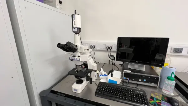
Leica DM4B Upright, motorised fluorescence/Brightfield microscope
Magnification, numerical aperture and immersion
- 2.5x, 0.7, air
- 5x, 0.15, air
- 10x, 0.30, air
- 20x, 0.40, air
- 40x, 0.65, air
- 63x. 0.80, air
Other features
- live Imaging chamber – No
- DIC – No
- stage – Manual
- camera – CCD colour and monochrome
- filter sets – DAPI, FITC, TRITC
Leica DM5000B Upright, motorised fluorescence/brightfield microscope
Magnification, numerical aperture and immersion
- 10x, 0.25, air
- 20x, 0.5, air
- 40x, 0.75, air
- 63x, 1.75, oil
- 100x, oil
Other features
- live Imaging Chamber – no
- DIC - no
- stage - manual
- camera - CDD colour and monochrome
- filter sets - DAPI, FITC, TRITC