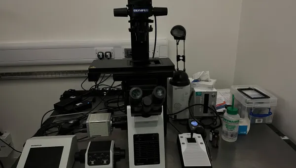
Live imaging system

More Information

About the Live imaging system

We have a live imaging system that allows us to investigate living cells using time lapse microscopy.

Live imaging is the processing of measuring dynamic physiological processes in living cells. This involves using specific fluorescence tags such as green fluorescent protein (GFP). 

The microscope is specially equipped to preserve optimal environmental conditions for the specimens. Data is usually collected as time-lapse movies.

Part of: Imaging and microscopy centre.

Technical specification

Deltavision Elite live imaging system
Modality and resolution
  • widefield deconvolution epifluorescence - 250 nm
  • DIC - 250 nm
Objectives
Magnification, numerical aperture and immersion
  • 10x, 0.5, air
  • 20x, 0.7, air
  • 40x, 1.4, oil
  • 60x, 1.42, oil
Specimen format
  • standard glass slide
  • 35 mm coverslip bottom dish
  • 40 mm coverslip bottom dish
  • coverslip bottom chamber-slide
Detector

1 x liquid cooled sCMOS. 2048 x 2048 pixels. 
 

Filtersets
Ex.Em.Channel
381-401 nm409-456 nmDAPI
400-454 nm463-487 nmCFP
425-495 nm500-550 nmGFP, FITC, Alexa 488
496-528 nm537-559 nmYFP
531-556 nm564-611 nmRhod, TRITC, Alexa 555
555-590 nm600-675 nmmCherry
619-644 nm652-700 nmCy5, Alexa 647
Other features
  • transmitted light - yes
  • DIC - yes
  • environmental temperature/gas control - yes
  • hardware autofocus - yes
  • simultaneous acquisition - no
  • sequential fluorescence acquisition - up to 4 channels
  • mark and find - yes
  • motorised stage with Tile Scan (stitch multiple FOV together) - yes
  • time-laspe - yes
  • photo-kinetics - no
  • 3D imaging - yes (with deconvolution)


Contact us

Dr Mark Willett

Highfield

Building 85, Highfield Campus, Southampton SO17 1BJ
We’re open Monday to Friday 09:00 to 17:00 UK time.
Back
to top